

The presence of 1qh+, 9qh+, inv(9)(p12q13), 15pstk+, or Yqh+ in a karyotype report is often lumped together as a “chromosomal abnormality.” According to cytogenetic nomenclature, these results mostly belong to common variations in chromosome morphology or heterochromatin regions; in clinical reports, the more accurate names are chromosomal polymorphisms or chromosomal heteromorphisms.
China's "Morphological Characteristics and Judgment Criteria for Human chromosomal polymorphisms and Expert Consensus on Genetic Counseling" released in 2026 systematically regulates common types, banding verification, tracing parental origin and genetic counseling. [1] The 2024 version of the Association for Clinical Genomic Science's karyotyping guidelines classifies well-documented benign abnormalities as normal variations, which are usually only recorded within the laboratory. [2] The two reporting strategies are different, but they share a common premise: confirm the exact morphology and breakpoint of the variant first, and then discuss the clinical significance.
This article discusses common polymorphisms in karyotyping reports. Aneuploidy, pathogenic deletions or duplications, reciprocal translocations, Robertsonian translocations, ring chromosomes, marker chromosomes, and definite mosaicism, with different genetic mechanisms and risk assessment pathways, do not fall into the low-risk category described here.
Read the karyotype first: what do 46, XX, p, q, and inv mean?
Taking 46,XX,inv(9)(p12q13) as an example, it can be understood in three sections:
- 46: 46 chromosomes were observed in the cells tested.
- XX: The sex chromosome composition is XX.
- inv(9)(p12q13): A chromosome 9 is broken at two positions, p12 and q13, and the segments between the two breakpoints are inverted and rejoined.
Chromosomes are divided into two arms by the centromere. The short arm is denoted p and the long arm is denoted q. Both region and band numbering increases from near the centromere toward the end of the chromosome. p12 means "short arm region 1, band 2"; q13 means "long arm region 1, band 3". [3,4]
The Chinese term "centromere" is pronounced zhuó. In the sentence "The inversion contains the centromere," "centromere" is a complete noun; it means that the inverted segment spans the centromere. inv is the abbreviation of English inversion, that is, inversion. If the inverted segment contains centromeres, it is called pericentric inversion; if it does not contain centromeres, it is called paracentric inversion.
Karyotype notation is a highly compressed positioning language. It explains what is seen under the microscope, but it cannot alone answer whether the variant is pathogenic or affects fertility, nor can it replace molecular-level gene or copy number testing.

The clinical significance of structural changes depends on the region
A chromosome is not a uniform line of DNA. Euchromatin usually contains many genes and has relatively high transcriptional activity; the regions near the centromere, secondary constrictions and some short arms are rich in repeated sequences and heterochromatin. Conventional G-banding karyotyping observes the banding morphology of chromosomes at a specific resolution.
Common polymorphisms often involve repetitive sequences, heterochromatin, satellites, or satellite stalks. Take 9qh+, for example, which represents a relative enlargement of the heterochromatin region on the long arm of chromosome 9. There is indeed a visible difference in length or amount of repetitive DNA here, so it would be inappropriate to generalize to "no increase in material at all." More precisely: no clear imbalance in known dose-sensitive euchromatic regions was shown.
When G-banding morphology is insufficient to distinguish polymorphisms from abnormalities, the laboratory can use supplementary banding or molecular testing: [1]
- C-band highlights the centromere and nearby heterochromatin and is commonly used to validate qh+, cenh+, and partial Y chromosome variants.
- The N band shows a transcriptionally active nucleolar organizing region, which can help determine whether the satellite stalk is extended or double.
- FISH, MLPA, CMA, CNV-seq, OGM or breakpoint sequencing can confirm origin, copy number or structure under specific questions.
Four common groups of polymorphisms in karyotype reports
1. Long-arm heterochromatin region variation: qh+ and qh−
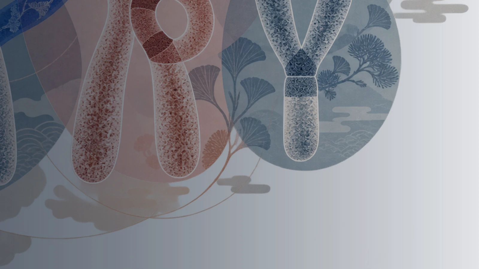
qh+ indicates a relative increase in the long-arm heterochromatin region, and qh− indicates a relative decrease. Commonly found on chromosomes 1, 9, and 16, but also found on the Y chromosome, such as 1qh+, 9qh+, 16qh+, Yqh+, Yqh−. [1,2]
This set of variations is primarily microscopic size or banding differences. The key points of judgment include: whether the variant is located in the expected heterochromatin region, whether the morphology conforms to the standard map, and whether it is consistent with the parent. Yqh− requires special caution because an excessively short Y chromosome may be confused with a deletion of the azoospermia factor region (AZF) of the Y chromosome; the 2026 Chinese consensus recommends combining C-banding and verifying the AZF gene or microdeletion. [1]
2. Satellite and satellite stalk variation of acrocentric chromosomes in group D/G
Chromosomes 13, 14, 15, 21, and 22 are acrocentric chromosomes. Their short arms contain satellites and satellite stalks. Common symbols include: [1]
- ps+/ps−: the short-arm satellite is enlarged/reduced;
- pstk+: the satellite stalk is elongated;
- pss or pstkstk: double satellites or double satellite stalks;
- cenh+/cenh−: Centromeric heterochromatin increases/decreases.
These morphologies sometimes approximate the appearance of Robertsonian translocations, derived chromosomes, or small marker chromosomes. An abbreviation in a report is not a substitute for the identification process. If necessary, C-band, N-band, FISH or copy number testing should be combined and the parental karyotyping should be checked.
3. Pericentric inversion of the heterochromatic region of chromosome 9: inv(9)(p12q13)
inv(9)(p12q13) is one of the more common pericentric inversions in karyotyping. The detection rate varies between different populations and laboratories, with multiple population studies reporting roughly around 1%-2%. [5] is listed as an pericentric inversion polymorphism in the heterochromatic region of chromosome 9 in the 2026 Chinese Consensus. [1]
The complete breakpoint must be preserved here. Inversion of chromosome 9 can also be written as inv(9)(p11q12), inv(9)(p11q13), inv(9)(p11q21), inv(9)(p13q13), etc. The common polymorphism specified by the latest Chinese consensus is inv(9)(p12q13); other breakpoint combinations cannot automatically apply the same conclusion. [1]
It is a balanced structural change: the orientation of the centromere-containing segment between two breakpoints changes, with no net copy number gain or loss in the conventional sense. Carriers are usually phenotypically normal. Systematic reviews and population studies have not yet concluded that it is clearly pathogenic. [5,6]
4. Other specific inversions and Y chromosome morphological variations
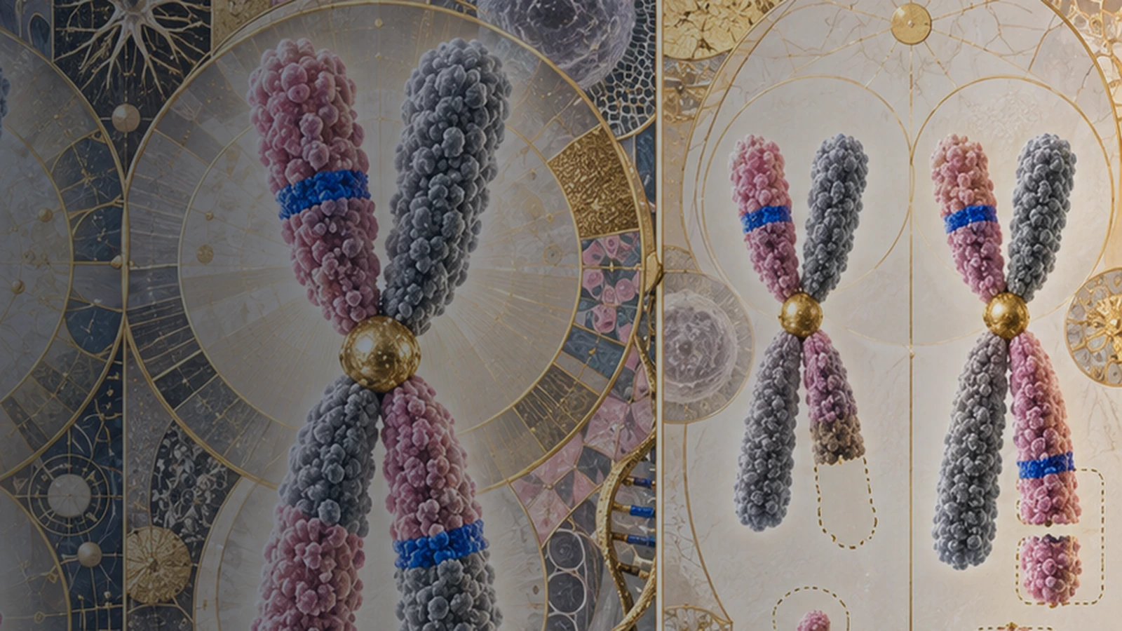
The 2024 Chinese PGT Genetic Counseling Consensus also lists in the example that “PGT-SR is not recommended only for common polymorphisms” inv(1)(p13q21), inv(2)(p11.2q13), inv(3)(p11.2q12), inv(10)(p11.2q21.2), inv(16)(p11.2q12.1), inv(Y)(p11.2q11.2) and Yqh+ etc. [7]
This list applies to exactly the same karyotype. When the breakpoint changes on the same chromosome, whether the inversion spans euchromatin, how many genes are involved, and how large the recombination fragment may be formed will all change accordingly. Clinical interpretation should be based on a complete karyotype and laboratory review.
Why do different guidelines provide different reporting methods?
The 2026 Chinese Consensus recommends describing the polymorphisms or variants seen in the report and providing explanations based on morphology, complementary banding, and parental origin. [1] The British ACGS 2024 guidelines regard benign abnormalities that have sufficient literature support as normal variations. In principle, they do not need to be included in patient reports, but laboratory records are retained. [2]
This difference mainly reflects laboratory reporting policies and genetic counseling culture, and does not mean that one system determines "disease" and another system determines "not disease". Both of them jointly emphasize:
- Benign aberrations need to be distinguished from pathogenic euchromatin deletions, duplications, or complex structural rearrangements;
- Special morphology, unexplained clinical phenotypes, and de novo variants require further investigation;
- The conclusion should indicate the detection method, resolution and limitations.
After getting the report, you should prioritize three questions: Has this pattern been accurately classified? Are there similar structural anomalies that need to be ruled out? How strong is the existing evidence to support clinical conclusions?
Existing reproductive and pregnancy research: overall signals and uncertainties
The answers given by existing studies are not entirely consistent because "chromosomal polymorphism" is not a homogeneous exposure factor. Different studies combine or split qh, satellite, Y variants and inversions, and the age of the study subjects, causes of infertility, IVF/ICSI methods, embryo culture and transplantation strategies are also different.
A 2023 systematic review and meta-analysis of 10 cohort studies found that female polymorphism carriers had an increased risk of miscarriage in pooled analyses, but pooled analyzes of male carriers or couples did not show the same association, and there were no consistent differences in outcomes such as clinical pregnancy, ongoing pregnancy, preterm birth, and live birth. [8] This suggests that there may be a signal in certain subgroups and also illustrates that observational data are susceptible to influence by the cause of infertility, age, and treatment choice.
A retrospective analysis of 11,739 infertile couples was conducted at Peking University Third Hospital in 2024. Overall, there was no stable overall difference in assisted reproductive outcomes between women with chromosomal polymorphisms and those with normal karyotypes, but associations in different directions emerged for different polymorphism types, IVF and ICSI subgroups. [9] A matched cohort study of male polymorphisms by the same team found that male inv(9) was associated with lower sperm motility, forward motility rate, and normal fertilization rate in the IVF subgroup, but no clear reduction in clinical pregnancy outcomes was found; there was an association with preterm birth in some small categories in the ICSI subgroup. The sample size and multiple comparisons require independent repeated verification of these results. [10]
A 2026 propensity score-matched cohort for Pericentric inversions of chromosome 9 that included 903 carrier cycles and 1,806 karyotype-normal control cycles. The number of oocytes retrieved, fertilization, blastocyst formation and most pregnancy outcomes were similar; there was no significant difference in the live birth rate of frozen embryo transfer, while the live birth rate of fresh embryo transfer was slightly higher in the carrier group. The study was single-center, retrospective, and autologous-oocyte, and “no disadvantage seen” cannot be interpreted as proof of zero risk for all populations and all breakpoints. [11]
Genetic pathways and unbalanced recombination of inv(9)(p12q13)
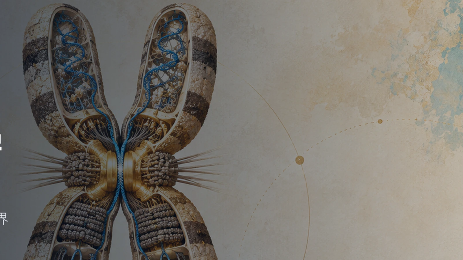
When one parent is a heterozygous inv(9)(p12q13) carrier and the other parent's two chromosome 9 structures are normal, each gamete will receive one of the two chromosome 9s. As far as chromosome transmission itself is concerned, the direct transmission of normal homologous chromosomes and inverted homologous chromosomes can be approximately understood as approximately half each.
This "about half" is the theoretical transmission probability. Each batch of embryos will fluctuate randomly, and fertilization, embryonic development and natural selection have not yet been taken into account. Judging from the inversion-related structure, the resulting embryo may have normal chromosome structure and carry the same balanced inversion as the parent, or in a few cases, unbalanced recombination related to inversions may occur.
pericentric inversions may form inversion loops during meiotic pairing. If exchange occurs within an inverted segment, recombinant chromosomes with distal duplications and deletions can theoretically be produced. The actual risk is affected by breakpoints, inverted-segment size, gender, and individual differences, and the average of all inversions cannot be used to replace the judgment of inv(9)(p12q13). In this small PGT series for this common inversion, no inversion-related imbalanced embryos were observed, but with a sample size of only 52 blastocysts, low-probability events cannot be excluded. [12]
If a fetus or child inherits the same inv(9)(p12q13) as a clinically normal parent, current consensus generally expects the clinical phenotype to be similar to that of the parent. [1] This conclusion relies on the premise of "consistent shape, clear source, and no other abnormalities". In an amniotic fluid cohort cited by the Chinese Consensus, 98.28% of the polymorphisms came from the parents, and no obvious growth and development abnormalities were found during follow-up to 1 year after birth; the follow-up time was short, so it is not suitable as an absolute guarantee of lifelong health. [1]
Detection boundaries for PGT-A, PGT-SR, CMA, and NIPT
The detection range depends on the technical principle. It is most likely to cause misunderstanding if all tests are collectively referred to as "chromosome testing".
PGT-A: Mainly assesses chromosome copy number in embryo biopsy samples
PGT-A typically analyzes chromosome copy number in trophectoderm biopsy cells using methods such as NGS or SNP. There is no net copy number change for balanced inversions, so conventional PGT-A generally cannot differentiate between "structurally normal" and "carrying the same balanced inversion" embryos. If the inversion recombination forms a duplication or deletion that is large enough and exceeds the laboratory's verified resolution, it may be detected. [13,14]
Therefore, “PGT-A euploid” should be understood to mean that no reported chromosome number or segment copy number abnormalities were found at this sample and platform resolution. It does not prove that chromosome orientation is completely normal, nor does it rule out all single-gene disorders, minor copy number variations, low proportions of mosaicism, or biopsy errors. [14,15]
PGT-SR: Applicability depends on structural anomalies and assay design
PGT-SR targets chromosome structural rearrangements. The 2024 Chinese Consensus clearly states that it is not recommended to implement PGT-SR only for common polymorphisms such as inv(9)(p12q13), 1qh+, 9qh+, Yqh+, etc. [7]
For true structural rearrangements requiring PGT-SR, common NGS or SNP copy number schemes may still fail to distinguish normal embryos from balanced-carrier embryos. The Chinese Consensus points out that many laboratories have limited diagnostic capabilities for CNVs below approximately 4 Mb on whole-genome amplification products of biopsy cells; before starting the project, it should be confirmed whether the expected imbalanced fragments are within the laboratory's validation range. [7] If the family goal is to distinguish between "normal" and "balanced carriage", breakpoint testing or haplotype linkage analysis may be required, and feasibility verification must be completed by the laboratory first. [13]
Karyotype, CMA and NIPT: different dimensions of observation
- G-banded karyotype can see larger numbers and structural changes, and can also identify balanced inversions; the resolution is limited and requires cultivable cells.
- CMA is good at detecting copy number gains and losses and generally cannot identify balanced inversions without net copy number changes.
- NIPT/cfDNA is a prenatal screening based on maternal plasma, not a fetal karyotyping diagnosis; routine projects cannot confirm balanced inversion, and positive or suspicious results require genetic counseling and diagnostic examinations.
- Karyotype analysis of chorionic villi, amniotic fluid or cord blood can directly observe whether the fetus carries balanced inversion; whether to add CMA should be decided based on ultrasound, family history and karyotype results.

Commonly used tests answer different questions
| Detection | Balanced inversions | Aneuploidy | Large deletions/duplications | Small CNVs |
|---|---|---|---|---|
| G-banding karyotype | Visible | Visible | Larger fragments visible | Usually limited by resolution |
| PGT-A | Usually cannot distinguish a normal karyotype from balanced-carrier status | Assessable | May be detected when verification threshold is reached | May be missed |
| Common CNV types PGT-SR | Usually unable to differentiate between normal and balanced carriage | Usually evaluable | Targeted evaluation | Often limited to ~4 Mb, subject to laboratory validation |
| CMA | Normally not visible | Visible | Visible | Depending on platform and probe coverage |
| NIPT/cfDNA | Unable to confirm | Screening only | Some changes may be screened | Not used as routine confirmation |
| Validated Breakpoint/Haplotype Scheme | Differentiable | Not Primary Use | Depends on the validated assay design | Depends on the validated assay design |
The American College of Obstetricians and Gynecologists emphasizes that there are false positives, false negatives, and non-coverage in preimplantation testing; routine prenatal screening or diagnostic testing should still be discussed after pregnancy. [15,16]
How to reach a well-supported conclusion after getting the "chromosomal polymorphism" report
Step 1: Check the complete karyotype
Confirm that the chromosome number, long and short arms, breakpoints and symbols are complete. inv(9)(p12q13) is not the same conclusion as other inv(9); 9qh+ is not the same change as inv(9).
Step 2: Confirm detection method and resolution
Check whether the report uses G banding or high-resolution banding, how many split phases were observed, and what the band resolution is. When the morphology is special, ask whether C-banding, N-banding or molecular verification has been done.
Step 3: Determine whether parental karyotyping is needed
When polymorphisms are discovered prenatally or in children, the parental karyotyping can determine whether they were inherited from clinically normal parents. For cases of new mutations, inconsistent morphology, or abnormal phenotypes, the normality of the parents does not end the evaluation, and additional testing still needs to be decided by clinical genetic professionals.
Step 4: Put phenotype and reproductive history back into the equation
When there are recurrent miscarriages, severe oligoasthenozoospermia, fetal structural abnormalities, developmental delay, or similar problems in the family, the scope of evaluation should cover other common causes. Attributing to one polymorphic karyotype alone can easily miss age, embryonic aneuploidy, uterine factors, endocrine factors, male factors or single gene diseases.
Step 5: Select testing according to specific reproductive path
Different information is required for natural conception, conventional IVF, PGT cycles and already pregnant cases. The need for PGT, prenatal diagnosis, and choice of karyotype or CMA should be determined jointly by the clinical geneticist, reproductive physician, and laboratory based on the specific karyotype and family goals.

Five sentences that can be directly used in report communication
- This result is a common chromosomal polymorphism identified by karyotype analysis, and its clinical significance requires a combination of complete karyotype and laboratory identification.
- Common polymorphisms generally do not equate to pathogenic deletions, duplications, or aneuploidies, but "common" does not constitute an guarantee of health for every individual.
- If the variant is morphologically consistent with the clinically normal parent, the expected clinical effects in the offspring are usually similar to those in the parent.
- PGT-A, CMA, or NIPT primarily detect copy number or screen risk and generally cannot reveal the balanced inversion itself.
- When new mutations, special morphology, abnormal phenotypes or adverse reproductive history occur, the karyotype should be reviewed and clinical genetic professionals should decide whether to supplement the test.
Conclusion
chromosomal polymorphisms are common phenomena in cytogenetics. Their core characteristics are that they are visible in human populations, have reproducibly identifiable morphological patterns, and most involve heterochromatin, satellites, or specific balanced inversions. Professional interpretation of such results requires respect for both morphological standards and evidence boundaries.
inv(9)(p12q13) is one of the most frequently asked examples. It is generally classified as a heterochromatic pericentric inversion polymorphism, existing assisted reproduction studies do not show a stable overall disadvantage, and conventional PGT-A does not recognize its balanced-carrier status. A complete conclusion should still retain three qualifications: the breakpoints must be consistent; similar structural abnormalities have been ruled out in the laboratory; individual phenotype, family history, and reproductive history do not suggest the need for expanded examinations.
Medical genetics needs to explain where the symbol comes from, what the test can see, how far the evidence supports it, and what questions still need to be addressed in specific families. Such hierarchical explanations are closer to real risks than a single “normal” or “abnormal” label.
References
Evidence search is open until September 20, 2026. The main sources are original research included in Chinese expert consensus, ACGS, ISCN, ESHRE, ASRM, ACOG and PubMed/PMC. This article is a popular science review and has not been reviewed systematically.
- [1] Weng Binghuan, et al. Morphological characteristics and judgment criteria of human chromosomal polymorphisms and expert consensus on genetic counseling. Heredity. 2026;48(1):3-25. DOI: 10.16288/j.yczz.25-146.
- [2] Association for Clinical Genomic Science. Best Practice Guidelines for Constitutional Karyotype Analysis and Targeted Chromosome Analysis. Version 1.0, 2024.
- [3] McGowan-Jordan J, Hastings RJ, Moore S, eds. ISCN 2024: An International System for Human Cytogenomic Nomenclature. Karger; 2024.
- [4] MedlinePlus Genetics. How do geneticists indicate the location of a gene? U.S. National Library of Medicine.
- [5] Šípek A Jr, et al. pericentric inversion of human chromosome 9 epidemiology study in Czech males and females. Folia Biol (Praha). 2015;61(4):140-146.
- [6] Mohsen-Pour N, et al. Chromosome 9 Inversion: Pathogenic or Benign? A Comprehensive Systematic Review of all Clinical Reports. Curr Mol Med. 2022;22(5):385-400.
- [7] Genetic Counseling Branch of the Chinese Genetics Society, et al. Genetic counseling expert consensus on preimplantation genetic testing. Chinese Journal of Obstetrics and Gynecology. 2024;59(12):899-909.
- [8] Ralapanawe MSB, et al. chromosomal polymorphisms in assisted reproduction: a systematic review and meta-analysis. Hum Fertil (Camb). 2023;26(3):687-698.
- [9] Lu Y, et al. Diverse impacts of female chromosomal polymorphisms on IVF/ICSI outcomes: a retrospective cohort study. BMC Pregnancy Childbirth. 2024;24.
- [10] Lu Y, et al. Impacts of male chromosomal polymorphisms on semen quality and IVF/ICSI outcomes: a retrospective cohort study. Int J Gynaecol Obstet. 2024;166(3):1247-1262.
- [11] Huang W, et al. Effect of pericentric inversion of chromosome 9 on reproductive outcomes in assisted reproductive technology: a propensity score-matched cohort study. Front Endocrinol. 2026;17:1811641.
- [12] Merrion K, et al. pericentric inversion (Inv) 9 variant—reproductive risk factor or benign finding? J Assist Reprod Genet. 2020;37:255-260.
- [13] Carvalho F, et al. ESHRE PGT Consortium good practice recommendations for the detection of structural and numerical chromosomal aberrations. Hum Reprod Open. 2020;2020(3):hoaa017.
- [14] Practice Committees of the American Society for Reproductive Medicine and the Society for Assisted Reproductive Technology. The use of preimplantation genetic testing for aneuploidy: a committee opinion. Fertil Steril. 2024.
- [15] American College of Obstetricians and Gynecologists. Preimplantation Genetic Testing. Committee Opinion No. 799. Obstet Gynecol. 2020.
- [16] American College of Obstetricians and Gynecologists. Ethical Considerations for Genetic Testing and Counseling in Obstetrics and Gynecology. Committee Statement. 2026.
Medical notice
This article is for public scientific education and does not substitute for clinical diagnosis, genetic counseling, or individualized medical advice. Karyotype interpretation should be completed by qualified cytogenetic laboratories and clinical genetic professionals based on original images, detection resolution, family history, and clinical manifestations.
Interpret the notation within the complete clinical picture
FS can help organise karyotypes, parental testing, reproductive history and reports from different laboratories. A clinical geneticist, fertility specialist and laboratory should determine individual risk and any additional testing.
Consensus, guidelines and research
The article preserves karyotype notation, breakpoints, study populations and technical resolution limits. A qualified cytogenetics laboratory and clinical-genetics professional should review individual reports.
- Chinese expert consensus on chromosomal polymorphisms (2026)
- ACGS karyotype-analysis guideline (2024)
- ISCN 2024
- MedlinePlus: cytogenetic location
- Population study of pericentric inversion 9
- Systematic review of chromosome 9 inversion
- Chinese PGT genetic-counselling consensus (2024)
- Meta-analysis of chromosomal polymorphisms in ART
- Female chromosomal polymorphisms and IVF/ICSI
- Male chromosomal polymorphisms and IVF/ICSI
- Matched ART cohort of inv(9) (2026)
- PGT study of inv(9) blastocysts
- ESHRE PGT technical recommendations
- ASRM committee opinion on PGT-A (2024)
- ACOG: Preimplantation Genetic Testing
- ACOG: Ethics of genetic testing and counselling (2026)
This article is for public science education and does not replace diagnosis, genetic counselling or individual medical advice.