

A balanced translocation is a structural chromosome rearrangement: genetic material has moved, but routine testing shows no obvious net gain or loss. Most carriers are healthy. The main consequences arise during gamete formation, embryo development and pregnancy.
Medication, supplements, diet or surgery cannot restore the chromosome arrangement. Clinical care instead reviews the karyotype and breakpoints, estimates individual risk, compares natural conception with PGT-SR and other pathways, and verifies the pregnancy with diagnostic testing.
1. A balanced translocation is not automatically a chromosome disease
Human somatic cells usually have 46 chromosomes. A reciprocal translocation exchanges segments between two non-homologous chromosomes and usually leaves 46 chromosomes. A Robertsonian translocation joins the long arms of two acrocentric chromosomes and often leaves 45. Balanced describes net copy number at the resolution tested; it does not mean normal position or risk-free gametes.
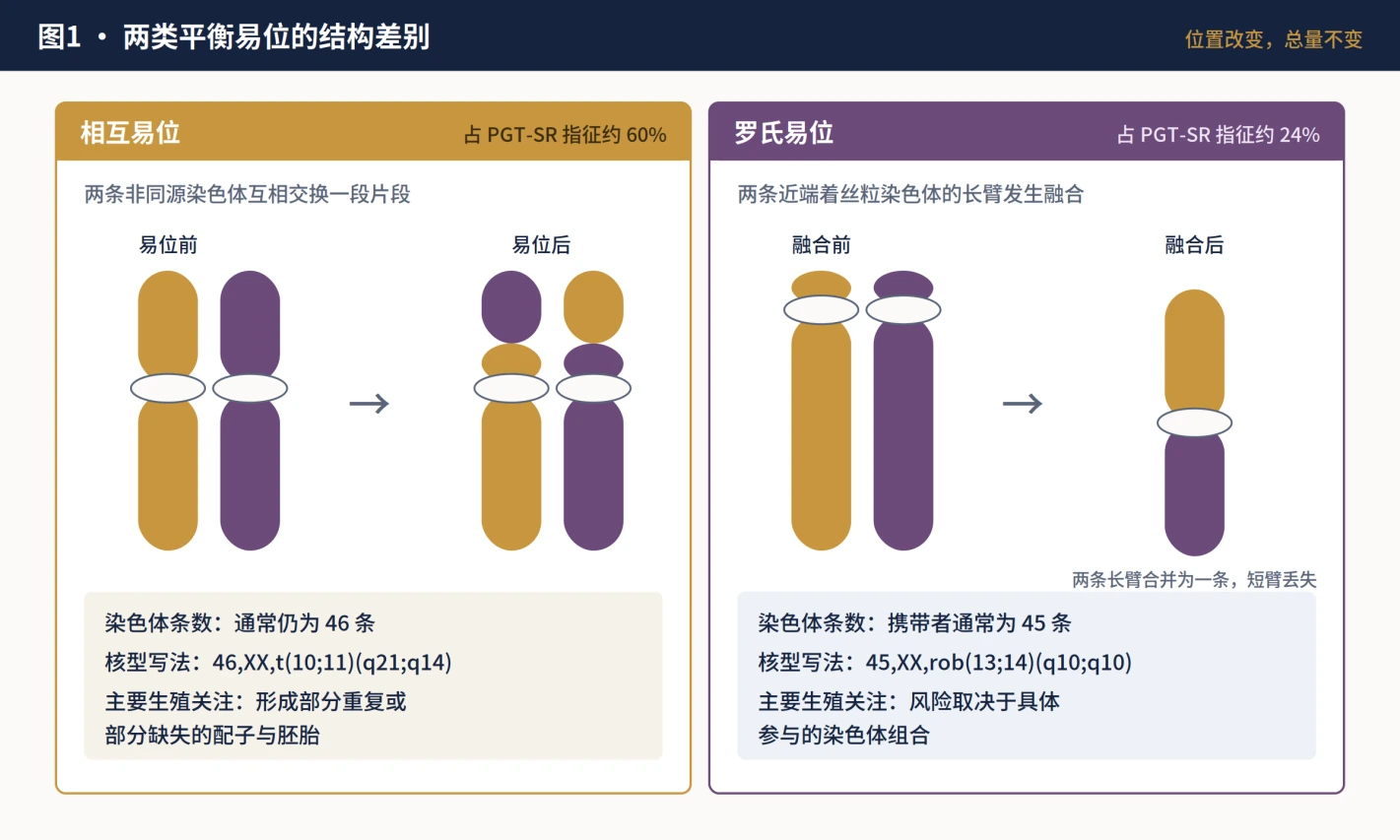
| Type | Structure and count | Main reproductive concern |
|---|---|---|
| Reciprocal | Segments exchanged; usually 46 chromosomes | Gametes and embryos may have partial duplication or deletion |
| Robertsonian | Long-arm fusion of two acrocentric chromosomes; usually 45 | Risk depends on the chromosomes involved; some combinations require UPD counselling |
Reciprocal translocations occur in roughly 1 in 500 people. A balanced structural rearrangement is found in about 2%-5% of couples with recurrent pregnancy loss. No universal percentage predicts an individual; type, breakpoints, chromosomes, carrier sex, maternal age and reproductive history all matter.
2. Testing order and karyotype interpretation
ASRM 2026 recommends chromosome testing of miscarriage tissue first when feasible, preferably with an array-based method. Parental blood karyotypes are most informative when miscarriage testing finds an unbalanced structural rearrangement or no miscarriage tissue result is available. ESHRE and RCOG also emphasize individual risk assessment.
About 50%-60% of first-trimester miscarriages are caused by sporadic embryonic aneuploidy and are strongly related to maternal age. Testing miscarriage tissue helps avoid assigning every loss to a parental karyotype. Once a balanced translocation is known, however, the result still guides counselling, pathway selection and assessment of at-risk relatives.
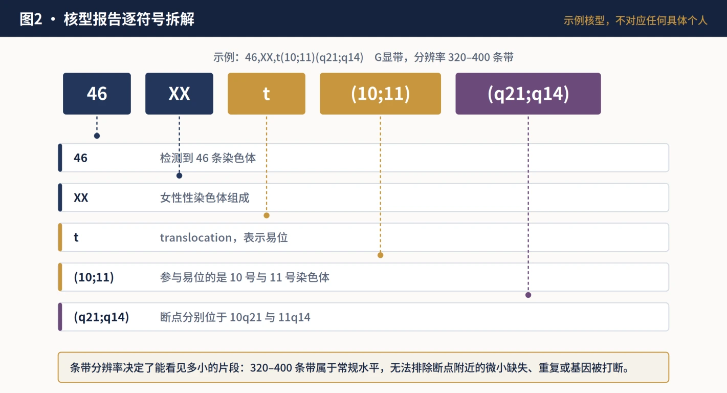
G-banding detects large patterns but can miss small copy-number changes or breakpoint complexity. An abnormal phenotype, complex family history or predicted segment near the platform limit warrants specialist review of higher-resolution karyotyping, FISH, microarray or breakpoint analysis.
3. A healthy carrier can still produce unbalanced gametes
During meiosis, the translocation chromosomes must pair and segregate. Broadly, gametes may be chromosomally normal, carry the same balanced rearrangement, or be unbalanced. Normal and balanced-carrier embryos can usually develop normally. Unbalanced embryos may arrest before biopsy, fail to implant, miscarry, or in a minority of cases continue with congenital or developmental consequences.
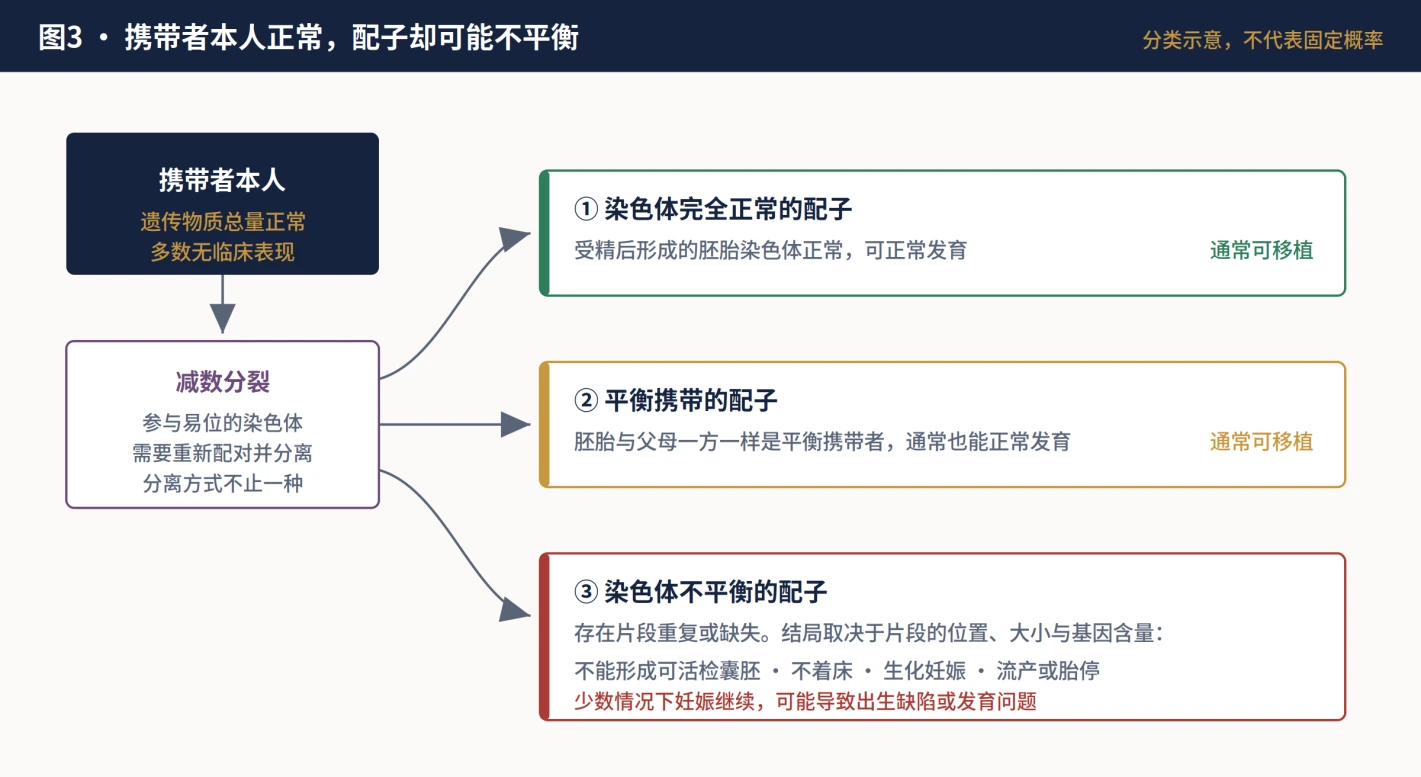

Translocations involving acrocentric chromosomes 13, 14, 15, 21 or 22 may have more 3:1 segregation. Rearrangements involving imprinted chromosomes 6, 7, 11, 14, 15 or 20 require discussion of uniparental disomy. Internet-wide fixed percentages are not individual counselling.
4. There is an association with IVF failure, but not automatic causation
Balanced translocations increase the proportion of unbalanced embryos and therefore have a biologically plausible relationship with blastocyst attrition, implantation failure and pregnancy loss. Two small studies cited by ASRM found unbalanced or other aneuploid results in 65.5% and 69.4% of embryos. Their size does not support individual prediction.
5. Natural conception and PGT-SR: similar destination, different burdens
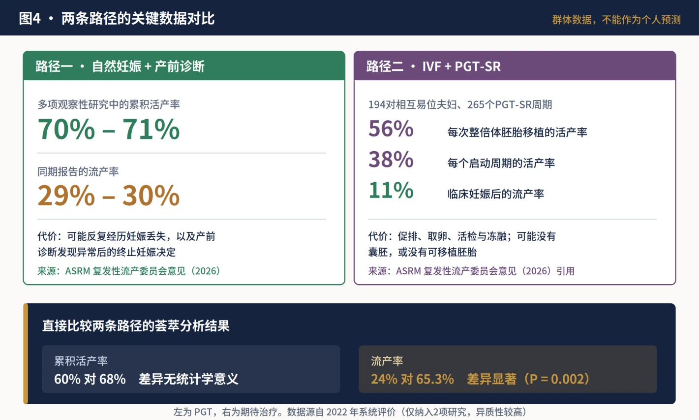
Natural conception followed by prenatal diagnosis is a guideline-recognized pathway. Observational studies summarized by ASRM report cumulative live-birth rates as high as 70%-71% without assisted reproduction and miscarriage rates around 29%-30%. These group results do not replace an individualized estimate.
IVF with PGT-SR aims to identify translocation-related imbalance before transfer. In 265 cycles among 194 reciprocal-translocation couples with adverse histories, live birth was 56% per euploid transfer, 38% per started cycle, and miscarriage 11% per clinical pregnancy. The denominators are fundamentally different.
| Pathway | Main value | Burden | Shared requirement |
|---|---|---|---|
| Natural conception + prenatal diagnosis | Avoids stimulation, retrieval and embryo biopsy | May involve repeated pregnancy loss and decisions after an abnormal prenatal result | Genetic counselling and diagnostic prenatal testing |
| IVF + PGT-SR | Reduces transfer of detected unbalanced embryos | May yield no blastocyst, no result or no transferable embryo; retrieval and cost burden | Prenatal diagnosis remains discussable |
| Donor egg or sperm | Can bypass the carrier gamete in selected cases | Access, legal, ethical and family implications | Local rules and counselling |
A 2022 systematic review found only two direct comparative studies. Cumulative live birth was 60% with PGD/PGT versus 68% with expectant management, without a significant difference; miscarriage was 24% versus 65.3%, lower with PGD/PGT. Sparse and heterogeneous evidence supports fewer losses, not a proven increase in cumulative live birth.
6. Ask for the denominator before discussing success
Per-biopsied-embryo rates answer the chance that an embryo yields a transferable result. Per-transfer rates exclude everyone who never obtained a transferable embryo. Per-started-cycle or per-analysis rates include no blastocyst, failed or inconclusive testing, and no transferable embryo.

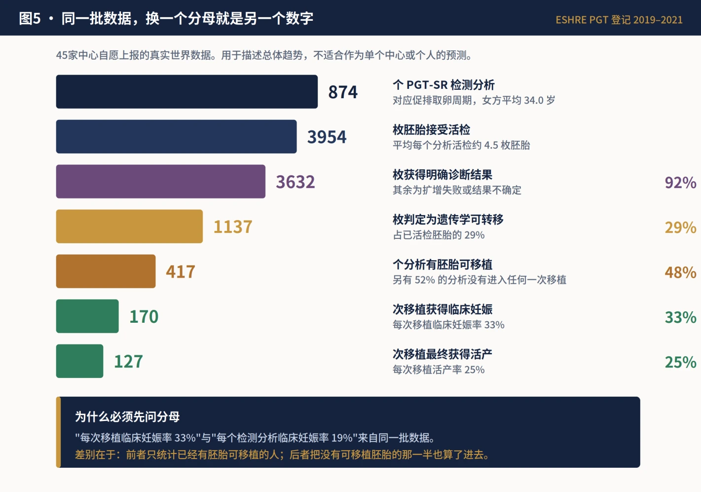
The ESHRE 2019-2021 registry included 874 voluntarily reported PGT-SR analyses from 45 centres. Diagnostic efficiency was 92%, but 52% of analyses had no transfer. Clinical pregnancy was 33% and live birth 25% per transfer; cumulative clinical pregnancy was 19% per analysis. These describe a registry, not a clinic or an individual prognosis.
A study of 1,942 PGT-SR cycles cited by ASRM found that roughly 4.5 biopsied blastocysts were needed, on average, for a good chance of at least one transferable embryo. Advanced age or diminished ovarian reserve may require multiple retrievals, so age, AMH/AFC and prior response should be modelled before treatment.
7. Confirm PGT-SR technical boundaries in writing
PGT-SR only works when the laboratory can detect the major imbalances expected from the specific rearrangement. Copy-number methods have finite resolution. A 2024 Chinese consensus notes that conventional NGS or SNP-array copy-number testing after embryo-cell amplification may not reliably detect segments around or below 4 Mb; the actual threshold depends on local validation.

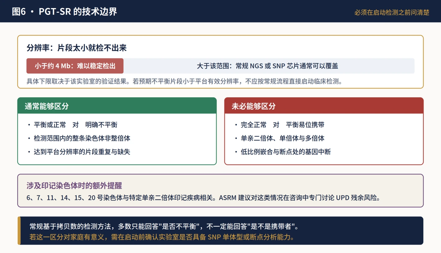
| Question before starting | Written answer needed |
|---|---|
| Was the karyotype reviewed? | Original karyograms, band resolution and any FISH or breakpoint confirmation |
| Are expected segments detectable? | Segment sizes, validated platform resolution, failure and no-result rate |
| Normal versus balanced carrier? | Many copy-number methods group them together; ask about haplotyping or breakpoint strategies if distinction matters |
| What remains outside scope? | Mosaicism, haploidy, polyploidy, UPD and other untested abnormalities |
| What if testing fails? | Policy for amplification failure, inconclusive result, re-biopsy and no transferable embryo |
8. PGT-SR is not the endpoint of fetal diagnosis
A blastocyst biopsy samples a few trophectoderm cells, which mainly form placenta, not the fetus. Amplification, mosaicism, sampling and platform resolution all matter. Chinese consensus and ACOG recommend that pregnancies after PGT still be offered prenatal diagnostic options.
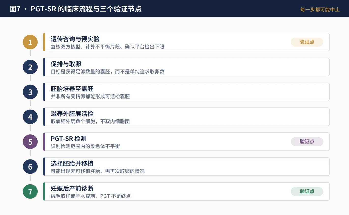
Chorionic-villus sampling mainly reflects placenta; amniocentesis samples fetal cells more directly. Timing, breakpoints and mosaic risk determine the test combination. Cell-free DNA screening is screening and cannot by itself replace invasive diagnosis targeted to a known translocation.
9. Put individual constraints into one decision table
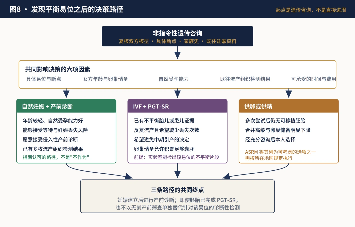
| Often supports discussion of natural conception | Often supports discussion of IVF + PGT-SR | Also discuss donor gametes |
|---|---|---|
| Younger age and good natural fertility | Known unbalanced fetus or substantial adverse history | Repeated PGT-SR with no transferable embryo |
| Accepts time and miscarriage risk | Priority is reducing repeated or later loss | Advanced age with markedly diminished reserve |
| Accepts diagnostic prenatal testing | Reserve is likely to support several blastocysts | Chosen after non-directive counselling |
No pathway is best for every carrier. Some people find another miscarriage hardest to bear; others find repeated retrievals with no embryo harder. Non-directive counselling should make probability, time, bodily burden, cost and decisions after an abnormal result visible.

10. Record checklist for the next consultation
| Step | Prepare or confirm |
|---|---|
| 1 | Original karyograms for both partners and specialist review |
| 2 | Family history of miscarriage, birth defects, developmental disorders and infertility |
| 3 | Prior retrieval, fertilization, blastocyst, transfer and embryo/miscarriage genetic records |
| 4 | Written laboratory feasibility, resolution, normal/carrier distinction and UPD scope |
| 5 | Age, AMH/AFC and prior response translated into expected blastocysts and acceptable retrievals |
| 6 | Compare natural conception, PGT-SR and donor gametes without success guarantees |
| 7 | Plan prenatal diagnosis after pregnancy; do not substitute NIPT alone |
Put the karyotype, breakpoints, ovarian reserve and laboratory feasibility in one review
FS helps families organize original karyotypes, prior embryo and pregnancy records, written laboratory feasibility and prenatal-diagnosis checkpoints. This article does not replace clinical genetics, reproductive medicine or prenatal-diagnosis care.
Guidelines and key studies
Core data and indications were checked against ASRM 2026, the 2026 ESHRE registry report, ESHRE/RCOG guidance, ACOG 799, the 2024 Chinese consensus and primary studies.
- ASRM Practice Committee. Recurrent pregnancy loss: a committee opinion, 2026.
- ASRM Practice Committee. Recurrent implantation failure: a committee opinion, 2026.
- Spinella F, et al. ESHRE PGT Consortium data collection XXII-XXIV: PGT analyses from 2019 to 2021, 2026.
- ESHRE PGT-SR/PGT-A Working Group. Good practice recommendations for detecting structural and numerical chromosome abnormalities, 2020.
- ESHRE PGT Consortium Steering Committee. Good practice recommendations for the organisation of PGT, 2020.
- ESHRE. Recurrent Pregnancy Loss Guideline, update 2022, published 2023.
- RCOG. Recurrent Miscarriage, Green-top Guideline No. 17, 2023.
- ACOG. Preimplantation Genetic Testing, Committee Opinion No. 799, 2020.
- Chinese expert consensus on genetic counselling for preimplantation genetic testing, 2024.
- Systematic review of subsequent pregnancy outcomes in couples with parental abnormal chromosomal karyotypes and recurrent pregnancy loss, 2022.
- Huang C, et al. Pregnancy outcomes before and after PGT-SR in reciprocal translocation carriers, 2019.
- Liu M, et al. Ovarian response and transferable embryos in balanced translocation carriers, 2022.
- Verdoni A, et al. Reproductive outcomes in individuals with reciprocal translocations, 2021.
Medical information only. Individual risk must be calculated by a clinical or reproductive genetics team using both karyotypes, breakpoints, age, ovarian reserve and prior pregnancy records.