FrankSense · Reproductive Genetics · Vol.110
Can PGT-M PreventMonogenic DiseaseTransmission?
PGT-M begins with the genetics of both partners.Inheritance pattern, carrier screening and family recordsdetermine whether a reliable test can be built.
Inheritance comes firstAD, AR, X-linked and mosaic risk models are different.
PGT-M reduces riskIt targets a known familial variant; it is not whole-genome clearance.
Confirmation still mattersCVS, amniocentesis or postnatal targeted testing should be discussed.

What PGT-M Is For
PGT-M, or preimplantation genetic testing for monogenic disease, is designed for families in which a specific pathogenic variant is already known. Its purpose is not to create a perfect embryo, but to reduce the chance that a defined inherited condition is passed to the next generation.
Whether PGT-M is appropriate cannot be judged from a carrier label alone. The key questions are whether both partners create a disease risk in the same gene pathway, how the condition is inherited, whether the variant has been confirmed by clinical testing, and whether the laboratory can build a dependable family-based assay.
This article assumes that the familial variant has been confirmed by clinical molecular testing, the relevant partner screening or family assessment has been completed, fertilization uses IVF/ICSI, and PGT-M is designed only for the known familial variant.
Inheritance first
01 | Inheritance Pattern Defines the Risk
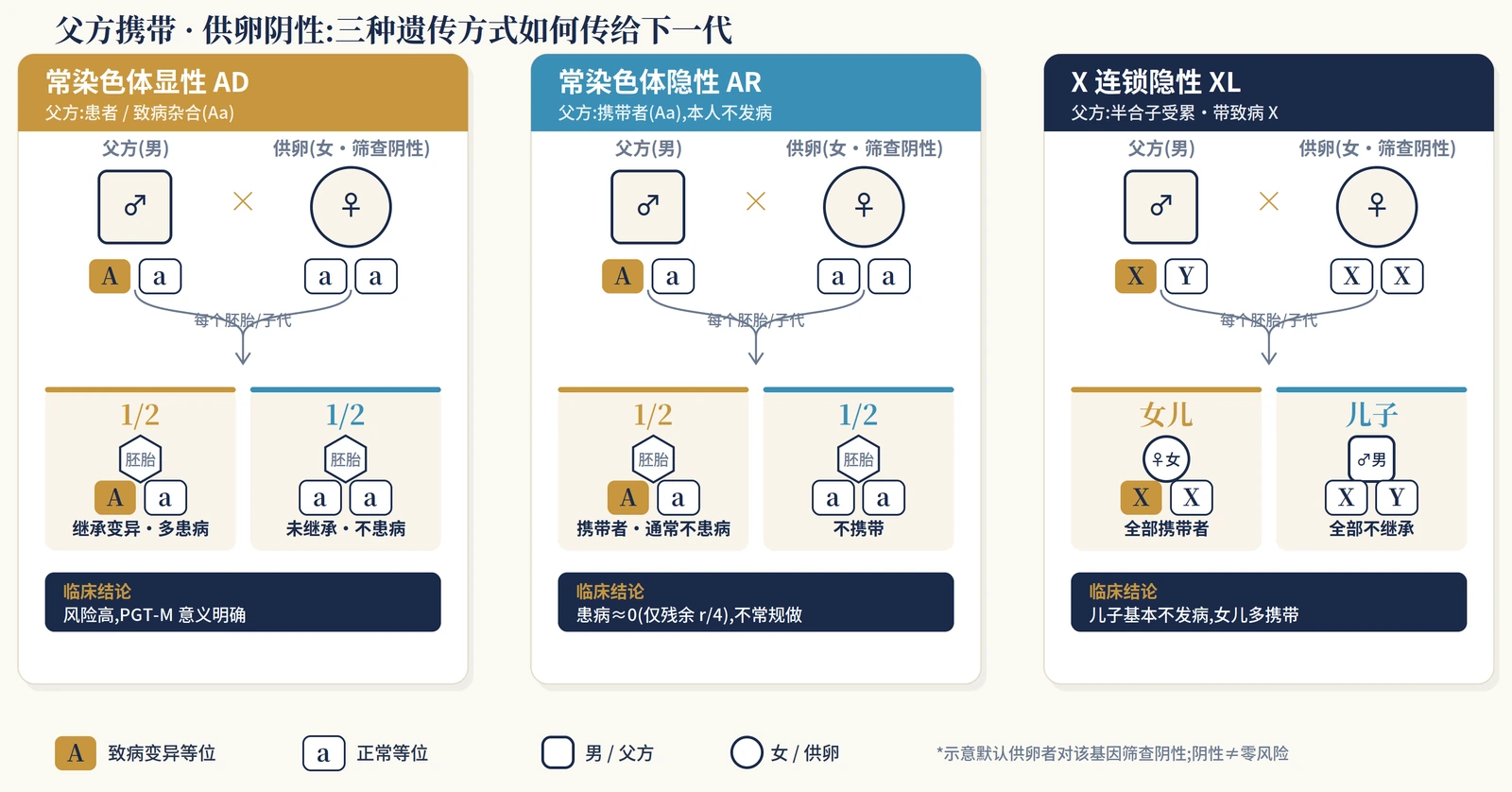
A carrier label is not precise enough for PGT-M counseling. The useful questions are: which gene is involved, which partner carries the variant, whether the other partner has risk in the same gene, how the condition is inherited, and whether embryo testing is clinically meaningful for this family.
The same familial variant can lead to very different counseling in autosomal dominant, autosomal recessive and X-linked disease. In practice, the discussion is closer to the table below than to a yes-or-no answer.
| Scenario | Assessment across both partners | Typical risk without PGT-M | Clinical relevance of PGT-M |
|---|---|---|---|
| Autosomal dominant | Either partner is heterozygous for a pathogenic variant, especially when personally affected or strongly suspected to be affected | Each embryo has about a 50% chance of inheriting the variant; penetrance and severity shape the actual disease burden | Clear risk, often a strong indication |
| Autosomal recessive | Whether both partners carry pathogenic variants in the same gene | When both partners carry the same-gene risk, each embryo is roughly 25% affected, 50% carrier and 25% unaffected; when only one partner carries, affected-child risk is usually much lower | Clear utility when both partners carry same-gene risk; usually not routine when only one partner is an asymptomatic carrier |
| X-linked recessive | Whether the female partner is a carrier, whether the male partner is affected, and whether the family wishes to avoid disease only or also carrier status | A carrier female partner may have affected male embryos and carrier female embryos; an affected male partner usually passes the X variant to female children but not to male children | Variant transmission and clinical disease must be explained separately |
| X-linked dominant | Which partner carries the variant, embryo sex where medically relevant, and disease severity | A carrier female partner may transmit risk to embryos of either sex; an affected male partner usually creates higher risk for female children and not for male children | Often clinically relevant, while sex information must follow local rules |
| De novo variant | Whether the variant is present in reproductive cells and whether it has been confirmed in the individual or family | If the variant is in the germline, transmission risk should not be dismissed merely because it first appeared as de novo | Requires molecular reports, family evidence and laboratory modelling |
| Gonadal mosaicism | Mosaic fraction, tested tissue, reproductive history and family evidence | Risk may be below the classic 50%, yet still far above population background risk | Individual modelling is required |
Note: the segregation rules for AD, AR and X-linked disease come from standard genetics; ASRM 2023 does not recommend routine PGT-M for asymptomatic one-partner AR carrier status; a negative screen still leaves residual risk depending on test scope and family background.
The most common mistake is to merge “inherits a variant” with “will develop disease.” Autosomal recessive carrier status, X-linked carrier status, reduced penetrance, mosaicism and de novo variants all need separate wording. Otherwise families may either overestimate risk because one variant appears on a report, or underestimate residual risk because a screen is negative.
PGT-M workflow
02 | What PGT-M Does: Reduce a Known Familial Risk
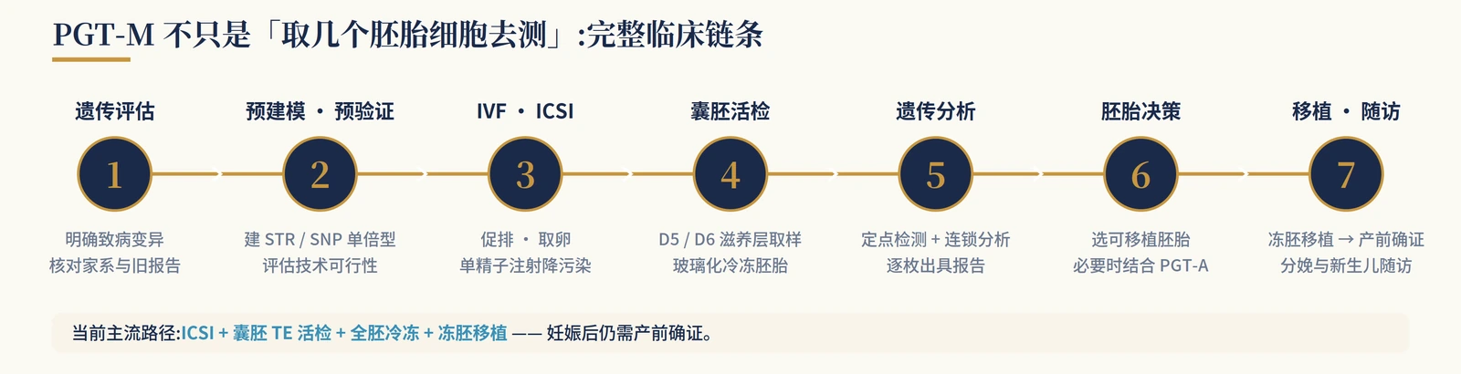
PGT-M is meant to reduce transmission of a known monogenic condition when the familial pathogenic variant is clear and a reliable assay can be built.
It is not whole-genome clearance and should not be marketed as a universal way to improve pregnancy rates. HFEA public information lists more than 2,000 approved genetic conditions for PGT-M in the UK, while ASRM notes that many Mendelian disorders with a known genetic cause may be considered, subject to feasibility, family samples and haplotype construction.
The clinical chain usually runs from genetic counseling and record review to family sample collection, haplotyping or linkage analysis, IVF/ICSI, blastocyst biopsy, genetic analysis, embryo decision-making, prenatal confirmation and newborn follow-up.
What should be prepared before a PGT-M cycle?
Before stimulation begins, the team usually needs the clinical diagnosis, molecular reports from both partners where relevant, evidence for variant pathogenicity, carrier screening results, family samples when needed, previous pregnancy or affected-child records, confirmation that informative markers can be built, and a check that the indication is permitted under local law and clinic policy. Better records make haplotyping easier; missing records may make the laboratory unwilling to issue a confident PGT-M plan.
Some cases are technically measurable but clinically unclear: variants of uncertain significance, very mild low-penetrance variants, one-partner autosomal recessive carrier status with a negative screen in the other partner, or families without enough samples for linkage analysis. Pushing forward in those settings may add cost, time and embryo loss without a clear clinical gain.
The technical point is straightforward: direct testing of the variant is usually stronger when combined with linkage analysis. STR or SNP haplotyping helps reduce errors from amplification failure, allele drop-out, contamination and recombination, especially when family samples are available.
| Method / combination | Principle | Typical use | Accuracy and limits |
|---|---|---|---|
| Direct mutation testing | Tests the known pathogenic site | Clear and technically simple variant | When used alone, more vulnerable to ADO and contamination; rarely ideal as the only evidence |
| Direct testing + STR/SNP linkage | Tests the variant and surrounding haplotype | One of the most common robust routes | Reduces target-locus misclassification; needs informative markers and often family samples |
| Karyomapping / SNP haplotyping | Genome-wide SNP-based haplotype construction | Family samples are available and a broader haplotype approach is useful | More robust against ADO and contamination; recombination still needs careful interpretation |
| WGA + targeted NGS / combined approaches | Amplify first, then perform targeted analysis | Comprehensive reporting after trophectoderm biopsy | Widely used; cannot eliminate mosaicism, recombination or sample identity errors |
| PGT-M + PGT-A | Adds aneuploidy information to PGT-M | Advanced maternal age, repeated failures or clinic-specific indications | More information, but not routine for everyone; may reduce the number of transferable embryos |
Note: “high accuracy” depends on case validation, family haplotype construction, laboratory experience and quality control. A promotional percentage should not be treated as a fixed value for every method and every center.
Reliability comes from variant confirmation, family modelling, linkage checks and laboratory quality control, not from a lone target-locus result.
Accuracy and residual risk
03 | High Accuracy Is Not Zero Risk
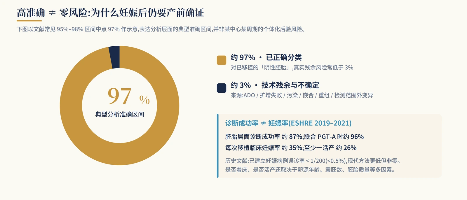
ACOG and ASRM both emphasize that false-positive and false-negative results can occur in PGT. A negative or unaffected PGT-M result does not rule out every genetic problem in a newborn, and it does not cover microdeletions, microduplications, de novo variants, imprinting disorders or conditions outside the designed assay.
How accurate is it? Published summaries and center-level reports vary, but a common broad range is about 95%-98%. For established pregnancies, historical misdiagnosis has been reported below 1/200 (<0.5%), and modern approaches may be lower. That is still not zero and does not remove the need for prenatal confirmation.
Residual risk can come from allele drop-out, amplification failure, contamination, embryo or placental mosaicism, meiotic or mitotic recombination, sample identity error, incorrect family information, and variants outside the tested region. Diagnostic success is also not the same as pregnancy rate or live-birth rate; implantation and live birth still depend on age, blastocyst number, embryo quality and the endometrium.
Should PGT-A be added?
The argument for adding PGT-A is stronger when maternal age is higher, prior cycles produced few usable embryos, or the goal is to avoid transferring embryos that are monogenic-variant-negative but clearly aneuploid. The cautious view is that ACOG and ASRM do not support PGT-A as a routine add-on for every IVF patient. Whether to add it should be judged by age, embryo number, prior cycles, clinic reporting policy and family goals. Mosaic embryo reporting and a smaller transferable embryo pool also need to be discussed before testing.
The better counseling is not “PGT-A is always necessary” or “PGT-A is useless.” It is to discuss age, testable blastocyst count, prior cycles, center policy and transfer goals in the same conversation.
Probability
04 | Probability: Embryo Number Changes the Chance
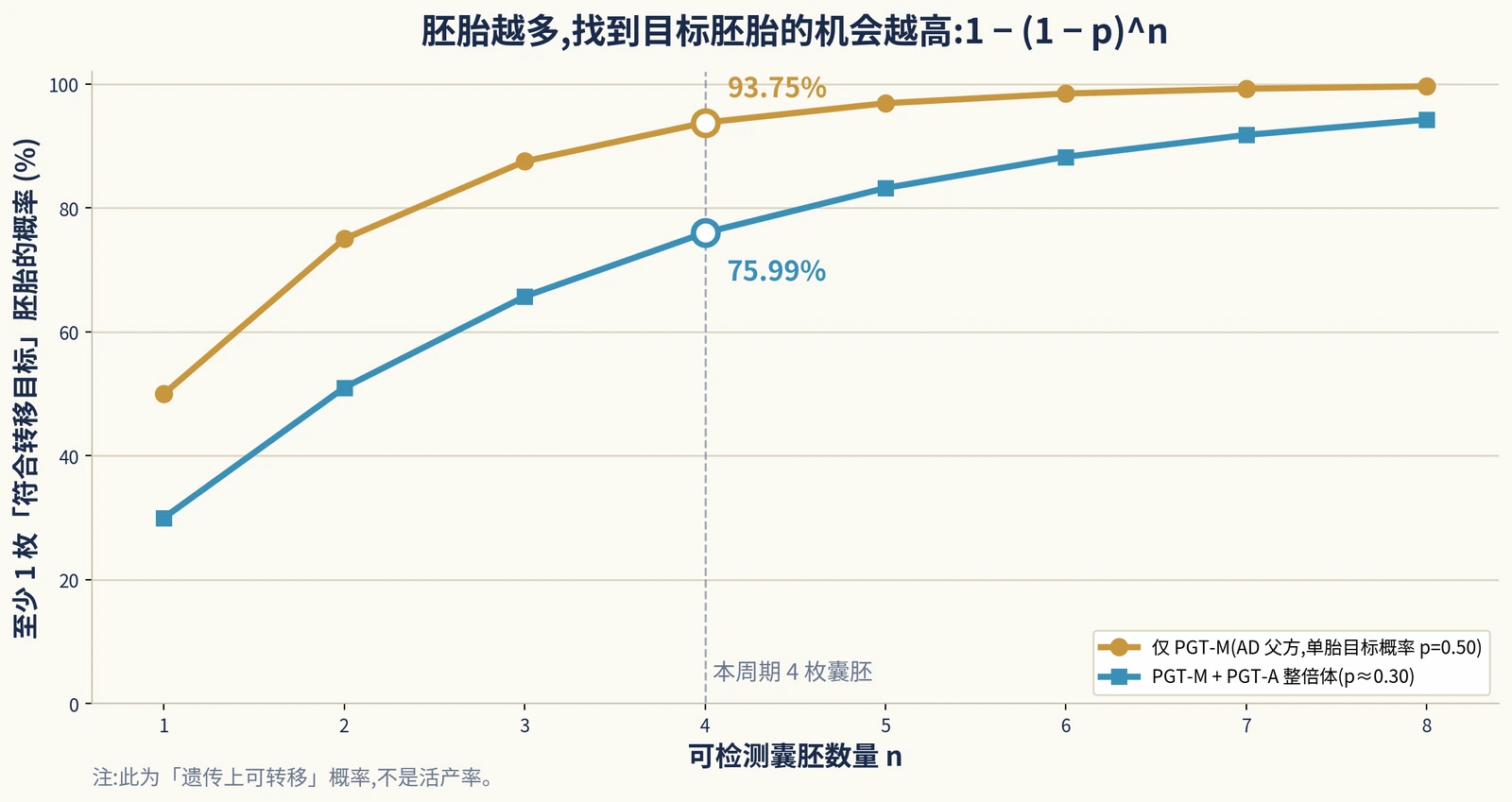
The practical formula is simple: each embryo has a probability p of meeting the transfer target; among n embryos, the probability of at least one target embryo is 1 - (1 - p)n.
Here, p may mean “does not carry a defined AD variant,” or it may mean “unaffected and euploid.” If euploidy is included, the extra assumption must be stated clearly. This is basic probability, not a live-birth promise.
Example 1: one partner carries an autosomal dominant variant
If one partner carries a high-penetrance autosomal dominant variant, each embryo has about a 1/2 chance of inheriting that variant and a 1/2 chance of not inheriting it. With four testable blastocysts, the chance of at least one embryo without the variant is 1 - 0.54 = 93.75%.
If euploidy is also part of the transfer goal, and if euploid probability is assumed to be about 60% and approximately independent of the monogenic locus, the chance of an embryo being both variant-free and euploid is about 0.5 × 0.6 = 0.30; four embryos give 1 - 0.74 = 75.99%. This shows that PGT-A changes the expected number of transferable embryos; it does not automatically improve live birth for every patient.
Clearer counseling: the embryo-level genetic risk for this condition is about 50%. PGT-M can substantially reduce that defined risk, but it is not a complete guarantee; prenatal confirmation should remain part of the plan.
Example 2: both partners carry the same autosomal recessive condition
When both partners carry pathogenic variants in the same gene, each embryo is roughly 25% affected, 50% carrier and 25% unaffected. If the family goal is to avoid affected embryos, about 75% of embryos may remain eligible for further discussion; if the goal is to avoid carrier status as well, the target group falls to about 25%.
This is why PGT-M counseling should define the transfer target early. Avoiding severe disease and avoiding carrier status are not the same goal. The former is often a clear clinical indication; the latter may raise more ethical, embryo-number and policy questions.
X-linked disease: carrier status and disease must be separated
X-linked disease cannot be summarized as “will it be inherited?” If the female partner carries an X-linked recessive variant, male embryos may be affected and female embryos may be carriers. If the male partner is affected, female children usually inherit the relevant X variant and male children usually do not. A strategy designed to avoid typical disease may differ from one designed to avoid any carrier status.
Whenever embryo sex information is involved, local law and ethics review must be followed. It can be discussed when medically relevant to disease risk; non-medical sex selection should not be presented as routine PGT-M.
One-partner AR carrier status and mosaicism need restraint
If only one partner is an autosomal recessive carrier and the other partner screens negative for the same gene, affected-child risk is usually already low. That is one reason ASRM 2023 lists asymptomatic one-partner AR carrier status as a non-recommended PGT-M indication. The real counseling need is residual-risk explanation, not pushing every carrier result toward embryo testing.
When gonadal mosaicism is suspected or proven, risk should be estimated from the mosaic fraction and reproductive history. Good counseling does not call it simply “low risk” or force it back to 50%; it places the evidence, test limits and reproductive options on the same page.
Ethics, law and cost
05 | Ethics, Law and Cost: Feasible Does Not Always Mean Appropriate

The strongest ethical indication for PGT-M remains serious, clear and verifiable monogenic disease. Its use has expanded into adult-onset disease, reduced penetrance and HLA matching, where the question shifts from “can it be done” to “should it be done.” Non-directive genetic counseling, patient autonomy and parallel discussion of alternatives are central.
Whether to transfer an embryo classified as affected is not answered by a slogan. It touches reproductive autonomy, the welfare of the future child, professional conscience and family context, and it requires clinic policy, informed consent and case-by-case judgment.
Regulation varies. The United States does not have a unified national PGT-M regulator, while the UK HFEA requires approved conditions and licensed clinics. Sex information is generally limited to medical indications in many jurisdictions.
| Cost item | Typical range | Comment |
|---|---|---|
| IVF / ICSI cycle | US about USD 15,000-25,000+; UK private about GBP 4,890-5,520 | Medication, region and lab package vary |
| PGT-M lab fee | US about USD 7,000-12,000 / cycle; UK private often from GBP 3,875 | Usually higher than PGT-A because custom test development is needed |
| Embryo biopsy / added analysis | Hundreds to thousands of dollars or pounds | Bundling and per-embryo pricing vary |
| PGT-A add-on | About USD 2,000-5,000+ | Should not be assumed for everyone |
| Frozen embryo transfer | UK about GBP 1,800-2,000; often separate in the US | Many PGT-M cases require freezing while results are pending |
Note: these figures are budget references, not contract terms. Insurance coverage, prior authorization and cross-region referral can materially change personal cost.
Timing is also longer than a standard IVF cycle. Genetic counseling and test development come first, followed by stimulation, biopsy, results and frozen embryo transfer. First-round commercial test development may take about 3-16 weeks; later cycles can be faster once the model is built.
Pregnancy and follow-up
06 | Pregnancy and Newborn Follow-Up

Once pregnancy is established, prenatal diagnosis should still be offered. ASRM, ESHRE and ACOG are aligned on this point: pregnancy after PGT-M should include counseling about rare misdiagnosis and the option of CVS or amniocentesis. If PGT-A was not performed, routine fetal aneuploidy screening or diagnosis should still be offered.
Follow-up should not stop at birth. If prenatal confirmation was not performed, targeted molecular testing after birth may be valuable. For X-linked disease, reduced penetrance, late-onset disease or clinically meaningful carrier status, family management and pediatric or specialty follow-up should be planned.
Current follow-up data are broadly reassuring, but should not be overstated. Available studies have not shown a major congenital malformation rate higher than IVF/ICSI controls, yet long-term outcomes by disease and testing pathway still require continued follow-up.
Responsible care links counseling, test development, embryo decisions, prenatal confirmation and newborn follow-up; it does not end on transfer day.
Conclusion
07 | Placing PGT-M in Real Decision-Making
Whether PGT-M is appropriate for a family depends on whether both partners' genetic information creates a defined disease risk, whether the inheritance pattern is clear, whether the variant is worth testing at the embryo level, and whether the laboratory can build a reliable assay.
The easiest error is to confuse “only one partner carries an AR variant” with “both partners carry same-gene AR risk,” or to treat “carries a variant” as the same as “will definitely be affected.” The former may require only residual-risk counseling; the latter may create a clear PGT-M indication.
PGT-M can substantially reduce transmission of a known familial variant, but it is not a total guarantee against all genetic, chromosomal, pregnancy or newborn risks. Its value is greatest when the indication, test boundary and pregnancy confirmation plan are stated plainly.
FAQ
Will a child definitely be affected if one partner carries a monogenic variant?
No. The answer depends on inheritance pattern, the other partner's genetic status, penetrance and whether the variant is present in the germline.
Can PGT-M guarantee a child without genetic disease?
No. It targets a known familial variant and still has residual risk.
Is PGT-M needed when only one partner is an autosomal recessive carrier?
Usually not if the other partner screens negative for the same gene; ASRM lists asymptomatic, one-partner AR carrier status as a non-recommended indication.
Is prenatal diagnosis still needed after PGT-M?
Patients should be offered prenatal confirmation by CVS or amniocentesis where appropriate.
Guidelines and Sources
This page summarizes public guidelines, regulatory information and professional recommendations for patient education and counseling.
- 胚胎植入前遗传学检测的遗传咨询专家共识. 中华医学会围产医学分会. 2025.
- 单基因病胚胎着床前遗传学检测专家共识. 中国医师协会生殖医学专业委员会 / 医学遗传医师分会. 2021.
- 胚胎植入前遗传学诊断 / 筛查技术专家共识. 2018.
- 血友病的胚胎着床前遗传学检测专家共识. 2021.
- ASRM Practice Committee: Indications and management of PGT-M, 2023 Source
- ESHRE PGT Consortium good practice recommendations for PGT-M, 2020 Source
- HFEA: Approved PGT-M and PTT conditions Source
- ACOG Committee Opinion No. 799: Preimplantation Genetic Testing Source
- ACOG: Carrier Screening in the Age of Genomic Medicine Source
- ACOG: Prenatal Genetic Testing Timing Source
- Hardy K, et al. Role of prenatal diagnosis after PGT; historical PGT-M misdiagnosis rate < 1/200. 2020.
- Poulton A, et al. Review: PGT-M accuracy approximately 95%-98%. 2025.
- Spinella F, et al. ESHRE PGT Consortium data 2019-2021.
- De Rycke M. PGT-M status review: technical evolution and offspring follow-up. 2020.
Turn a genetic report into a practical pathway
Bring both partners' reports, inheritance pattern, family samples, carrier screening and embryo count into one PGT-M feasibility review.
Review the pathwayThis article is educational and does not replace personal medical care, genetic counseling or laboratory-specific test review.