
Monochorionic diamniotic, or MCDA, is not another way of saying that one embryo was transferred and two babies appeared. It is an ultrasound-defined anatomy: two fetuses share one chorionic and placental circulation, while each remains in a separate amniotic sac. That anatomy sets the surveillance interval, warning signs and referral urgency.
Most MCDA twins result from one fertilized egg or embryo splitting and are therefore usually monozygotic. Chorionicity and zygosity are nevertheless different dimensions. Rare exceptions, fused placentas and classification error exist, so pregnancy care follows chorionicity while genetic zygosity can be tested separately when it matters.

1. Separate fetal number, zygosity, chorionicity and amnionicity
Fetal number asks how many fetuses are present. Zygosity asks how many fertilized eggs they came from. Chorionicity asks whether the placental system is shared. Amnionicity asks whether the amniotic cavity is shared. These questions are related but not interchangeable, and clinical risk is driven mainly by the last two.
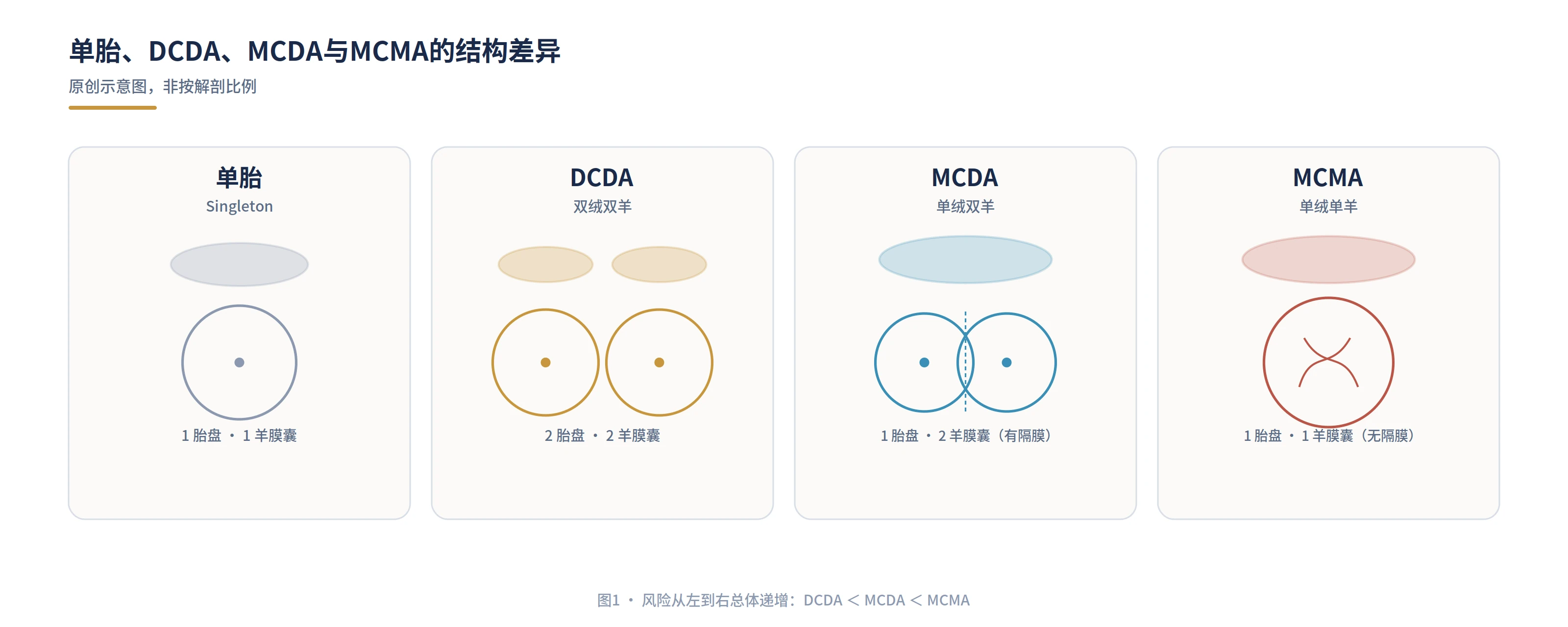
| Anatomy | Placental system | Amniotic sacs | Main added risk |
|---|---|---|---|
| DCDA | Two | Two | General twin risks including prematurity and growth complications |
| MCDA | One shared | Two | TTTS, TAPS, sFGR and acute co-twin injury through shared vessels |
| MCMA | One shared | One shared | MCDA risks plus cord entanglement and compression |
Dizygotic twins are almost always DCDA. Monozygotic twins may be DCDA, MCDA or MCMA. Dichorionic therefore does not mean dizygotic, and identical does not automatically mean MCDA. Sex, appearance and the number of placental masses cannot replace a standard ultrasound determination.
2. How twins form after spontaneous conception or embryo transfer
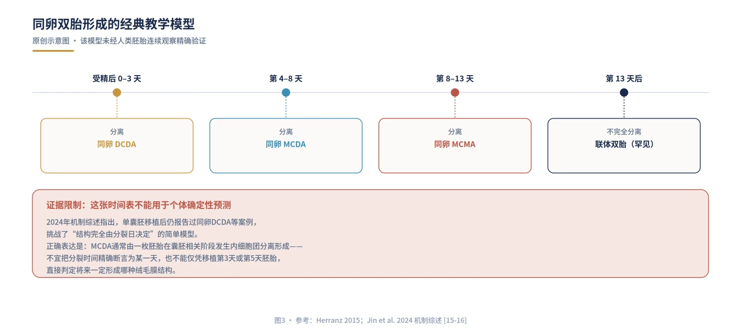
After spontaneous conception, two separately fertilized oocytes usually produce dizygotic DCDA twins. A single conceptus that divides can produce monozygotic DCDA, MCDA or MCMA twins. The classic timing model is useful for teaching, but current human evidence does not support using it as a precise calendar for an individual embryo.
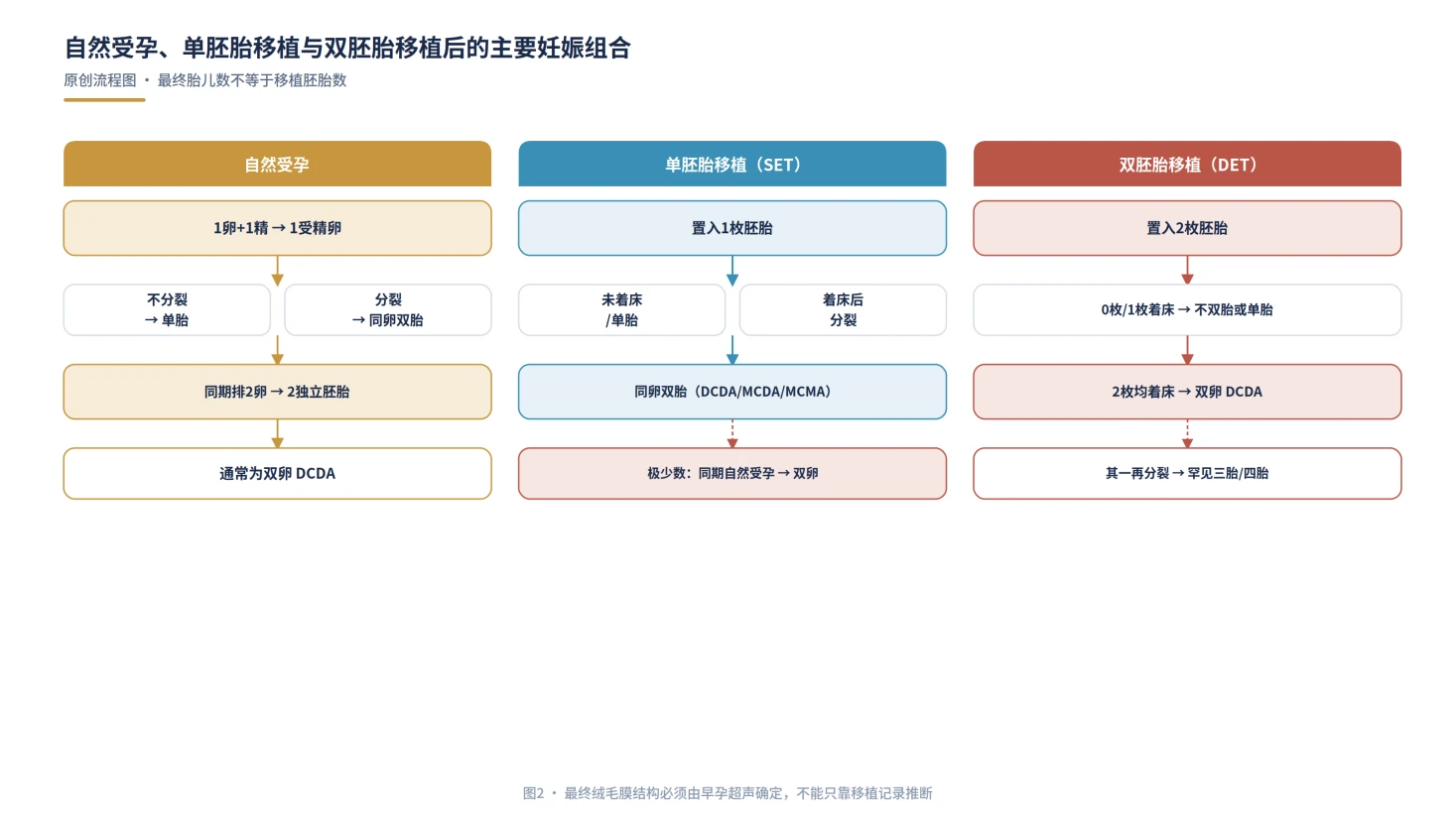
Elective single-embryo transfer greatly reduces treatment-related multiple pregnancy, but it does not make the risk zero. One embryo may divide. In a natural or stimulated frozen-transfer cycle, concurrent ovulation and intercourse may rarely add a spontaneously conceived embryo. Double-embryo transfer increases twins and can produce triplets if one embryo also divides.


ESHRE and ASRM frame the goal as one healthy term live birth at a time. For suitable patients, elective single-embryo transfer is the central strategy for reducing prematurity, low birth weight, NICU admission and maternal complications.
3. Why MCDA is a high-risk pregnancy
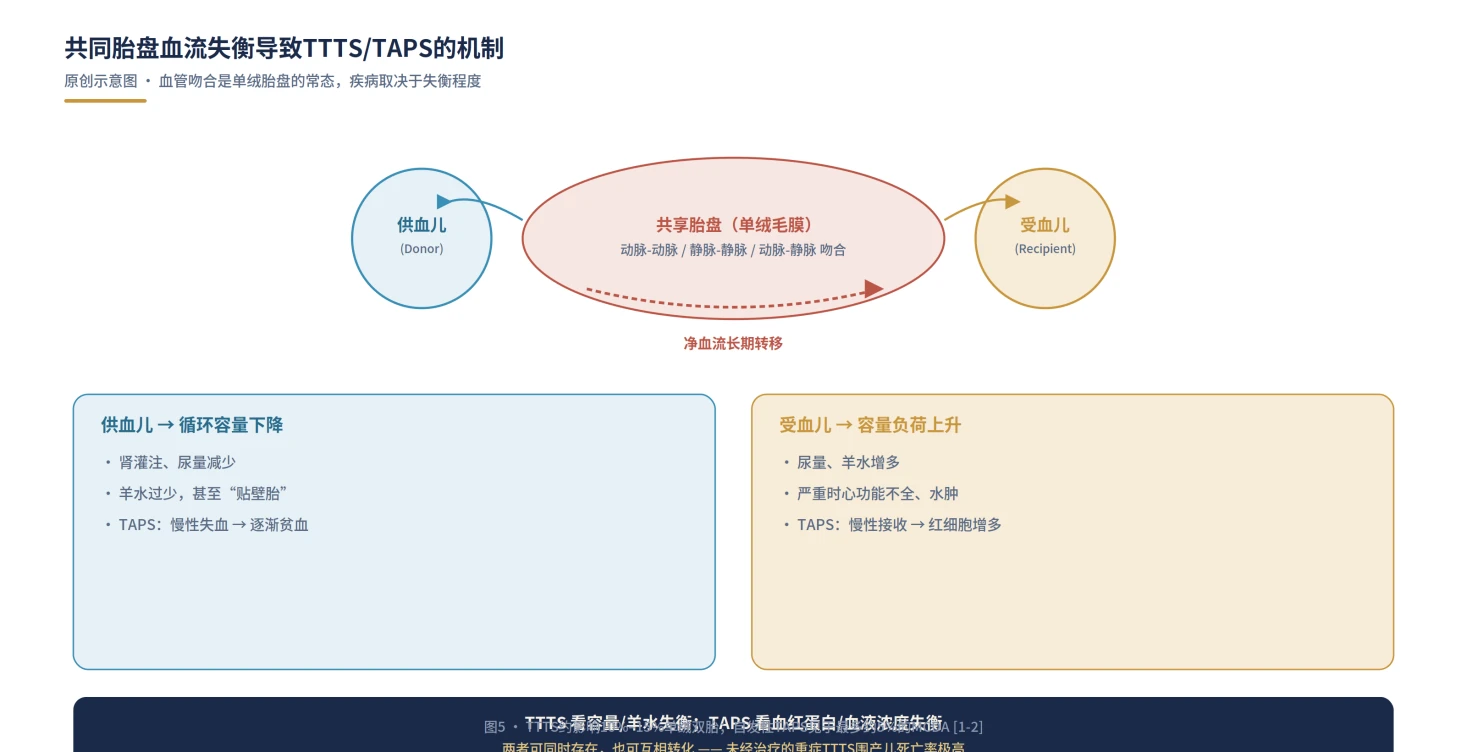
The defining hazard is not that two babies are genetically alike. A shared placenta usually contains vascular anastomoses connecting the umbilical circulations. These connections are common; disease depends on their number, direction, resistance and whether unequal placental territories can remain haemodynamically balanced.

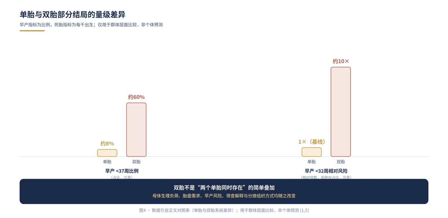
Twin pregnancy also increases uterine distension, cervical load, membrane rupture, anaemia, hypertensive disorders, caesarean birth and postpartum haemorrhage. Roughly six in ten twin pregnancies deliver before 37 weeks, and very preterm birth is much more common than in singleton pregnancy. Shared-circulation disease adds another layer, so MCDA cannot follow a routine low-risk twin schedule.
4. Four condition groups that must be recognized early
TTTS affects about 10%–15% of monochorionic twin pregnancies. The donor develops reduced circulating volume, urine output and amniotic fluid; the recipient develops volume loading, polyuria and excess fluid and may progress to cardiac failure. Diagnosis is based on an oligohydramnios-polyhydramnios sequence, not a weight difference.
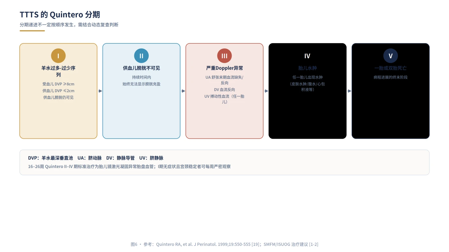
SMFM and ISUOG recommend fetoscopic laser ablation of abnormal placental vessels as standard treatment for Quintero stage II–IV disease between 16 and 26 weeks. Asymptomatic stage I disease with a stable cervix may undergo at least weekly surveillance, with laser considered when symptoms, progression or additional risk factors appear. Suspected TTTS should not wait several routine-visit intervals.
TAPS results from slow transfusion through very small anastomoses. One twin becomes anaemic and the other polycythaemic, often without the fluid discordance of TTTS. Prenatal screening relies mainly on the two MCA peak systolic velocities. Thresholds and starting gestation differ across guidance, so a fetal-medicine team must interpret the whole pattern.
sFGR usually reflects unequal placental territory. It is not enough to label one baby constitutionally small. Estimated-weight centiles and discordance, growth trajectory, and umbilical-artery and ductus-venosus Doppler all matter. Gratacós types I, II and III are defined by end-diastolic flow in the smaller twin's umbilical artery and have different prognoses.

When one MCDA twin dies, shared vessels may allow acute blood loss from the survivor into the low-resistance circulation of the deceased twin. Anaemia, cerebral ischaemia or co-twin death can occur around the event. Reflex immediate extreme-preterm delivery may not reverse established injury; rapid specialist assessment of MCA, Doppler, heart rate and brain imaging is needed.
5. Determine chorionicity by 11–13+6 weeks and preserve twin identity
Earlier determination is more reliable. The number of placental masses, membrane thickness and the lambda or T sign should be assessed together, and an image of the membrane-placenta junction retained. Two placentas may fuse, so one apparent placental mass is not enough. If expert review still cannot exclude monochorionicity, management should remain on the monochorionic pathway.
| Ultrasound feature | More consistent with DCDA | More consistent with MCDA |
|---|---|---|
| Membrane-placenta junction | Complete lambda or twin-peak sign | T sign or empty lambda |
| Intertwin membrane | Thicker, including two chorionic layers | Thin, mainly two amniotic layers |
| Placental appearance | Often two, but they may fuse | Usually one, but two masses may occasionally appear |
Twin A and Twin B should be mapped using several stable features such as maternal left or right, upper or lower position, placental position and cord insertion. If one twin has an anomaly or receives treatment, identity must be reconfirmed before birth because delivery order may not match antenatal labels.
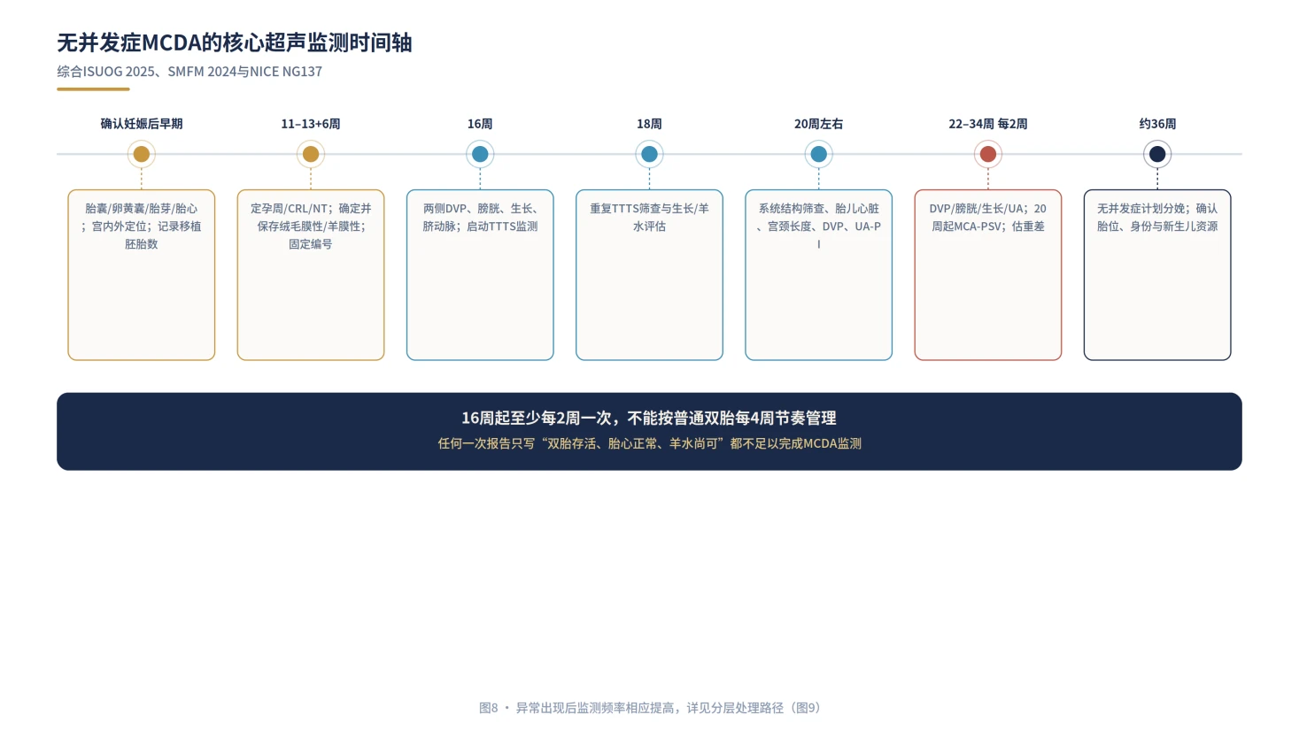
6. Uncomplicated MCDA: ultrasound at least every two weeks from 16 weeks
ISUOG, SMFM and NICE converge on a dedicated surveillance pathway beginning at 16 weeks and continuing until birth. Each visit should separately document the deepest vertical pocket, bladder, growth and umbilical-artery assessment for both twins. MCA-PSV is usually added from 20 weeks for TAPS screening; SMFM supports considering it from 16 weeks.

| Stage | Core assessment | Purpose |
|---|---|---|
| 11–13+6 weeks | Dating, NT, early anatomy, chorionicity, amnionicity and labels | Build the pregnancy risk and identity map |
| Every 2 weeks from 16 | DVP, bladder, growth and UA Doppler for both twins | Screen for TTTS and growth disease |
| Around 20 weeks | Detailed anatomy, heart, cervix, UA and MCA-PSV | Screen for anomalies, prematurity, TAPS and sFGR |
| After an abnormality | Weekly or more frequent specialist assessment | Guide fetal therapy or delivery by condition and stage |
A report stating only that both twins are alive with normal heartbeats and acceptable fluid is not an adequate MCDA record. Twin-specific measurements are required. Detailed anatomy at 18–22 weeks should be performed by an experienced twin operator, with particular attention to fetal cardiac assessment.
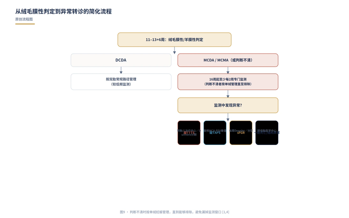
7. An abnormal finding immediately changes frequency and referral
| Finding | Immediate action | Next focus |
|---|---|---|
| Suspected TTTS | Contact a centre with fetoscopic laser capability | Quintero stage, cervix and cardiac assessment |
| Suspected TAPS | Repeat both MCA-PSV values and their discordance | Observation, transfusion, laser or delivery by stage and gestation |
| sFGR | At least weekly Doppler and classify type I, II or III | Balance small-twin death, co-twin brain injury and extreme prematurity |
| Death of one twin | Assess survivor anaemia, Doppler and brain imaging | Avoid unassessed reflex extreme-preterm delivery |
After fetoscopic laser, SMFM recommends weekly surveillance for six weeks followed by at least every two weeks, unless recurrent TTTS, post-laser TAPS or growth restriction requires more. When both twins survive and remain stable, delivery is commonly planned at 34–36 weeks in the absence of another indication.


8. Timing and mode of birth depend on anatomy, complications and resources
Uncomplicated MCDA pregnancy is usually offered planned birth at 36+0 to 36+6 weeks. MCMA is earlier, generally 32–34 weeks and usually by caesarean because of the shared sac and cord hazards. TTTS after laser, sFGR, TAPS or fetal compromise requires individualized timing.
MCDA does not mandate caesarean birth. Planned vaginal birth may be appropriate when the first twin is cephalic, gestation and estimated weights are suitable, there is no fetal compromise, and the unit can provide continuous dual monitoring, immediate caesarean capability and skilled management of the second twin.
9. ART, PGT and gestational-carrier implications
Final fetal number and placental anatomy cannot be reverse-engineered from the number of embryos transferred. After single-embryo transfer, two heartbeats most often reflect splitting, but rare concurrent natural conception remains possible. Early ultrasound should count sacs, yolk sacs and embryos and examine the adnexa for heterotopic pregnancy.
When one PGT-tested embryo divides, both twins usually inherit the original test result. But PGT samples a small number of trophectoderm cells; it does not replace prenatal ultrasound, screening or indicated diagnostic testing, and it cannot predict TTTS, TAPS or placental share.
Whether a gestational carrier supplied the oocyte does not determine zygosity and does not alter the shared-placenta risk. Carrier health shapes the overall obstetric risk, but it cannot remove the MCDA surveillance and referral requirements.
10. Seven answers the family and care team should retain
| Point | Required answer |
|---|---|
| Before transfer | Why SET or DET; intercourse and contraception instructions in natural cycles |
| First positive test | Timing of location scan; sac and embryo count; adnexal review |
| 11–13+6 weeks | DCDA, MCDA or MCMA; stored lambda/T-sign image; A/B labels |
| From 16 weeks | Dedicated two-weekly scans with separate DVP and bladder records |
| Around 20 weeks | Detailed anatomy, heart, cervix, UA, MCA and weight discordance |
| Any abnormality | Condition, severity and named fetal-therapy referral centre |
| Before and after birth | Timing, presentation, NICU, blood preparation, identity handover and neonatal checks |
Put chorionicity, gestation, twin identity and warning signs in one plan
FS helps families organize embryo-transfer records, early ultrasound, twin identity, surveillance timing and cross-border referral information. This article does not replace a fetal-medicine team.
Sources
Core recommendations were checked against ISUOG 2025, SMFM 2024, the RCOG 2024 partial update, NICE's 2024 update and ESHRE/ASRM guidance.
- ISUOG Practice Guidelines (updated): role of ultrasound in twin pregnancy, 2025.
- SMFM Consult Series #72: Twin-twin transfusion syndrome and twin anemia-polycythemia sequence, 2024.
- RCOG Green-top Guideline No. 51: Management of Monochorionic Twin Pregnancy, 2024 partial update.
- NICE NG137: Twin and triplet pregnancy, updated 2024.
- ESHRE Guideline: Number of embryos to transfer during IVF/ICSI.
- ASRM Committee Opinion: Multiple gestation associated with infertility therapy, 2022.
- Vitthala S, et al. Monozygotic twins after assisted reproductive technology: systematic review and meta-analysis.
- Hviid KVR, et al. Determinants of monozygotic twinning in ART: systematic review and meta-analysis.
- Jin H, et al. Cellular mechanisms of monozygotic twinning, 2024.
- Khalil A, et al. Consensus definition of selective fetal growth restriction in twin pregnancy.
- Gratacós E, et al. Classification of selective growth restriction in monochorionic twins.
Medical information only. MCDA abnormalities can evolve quickly. Fluid discordance, growth discordance, abnormal Doppler, maternal symptoms or death of one twin require immediate assessment by a team experienced in twin and fetal therapy.