
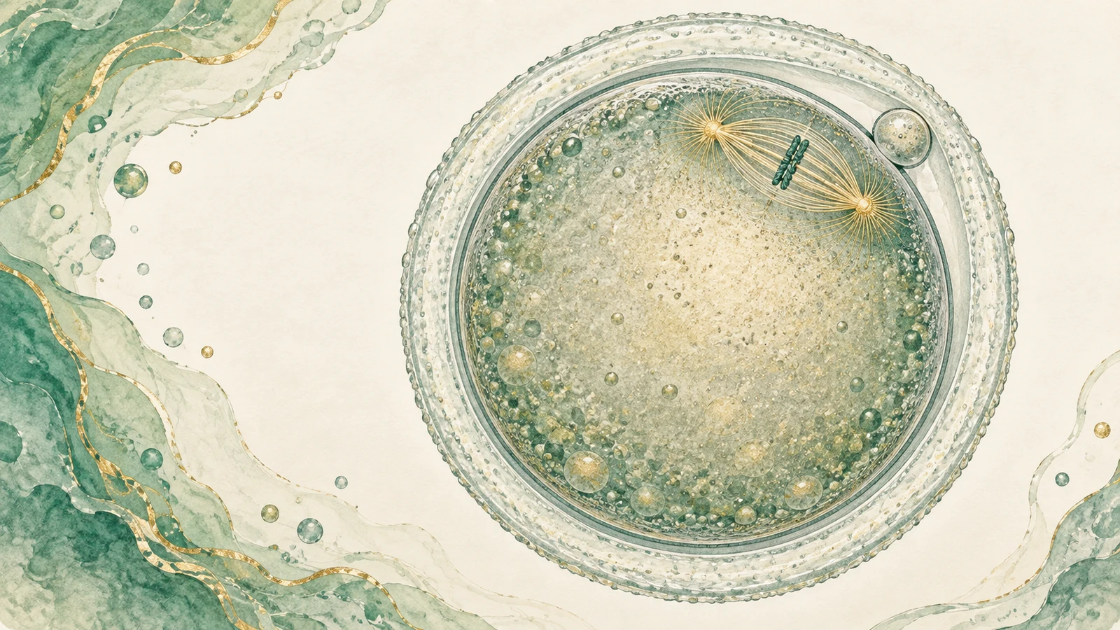
The laboratory determines whether the oocyte is mature based on whether the first polar body has been extruded. The Istanbul Consensus Update released by ESHRE and ALPHA in 2025 states that a portion of oocytes with the first polar body are still in telophase I, and the meiotic maturity status of the oocyte can only be determined by observing the metaphase II spindle in the cytoplasm. This raises the question of whether polarized-light spindle imaging can inform the timing of fertilization.
Current methods for determining oocyte maturation
The last step in the ovarian stimulation cycle is the trigger, which is the injection of a drug that prompts the oocyte to complete its final maturation, usually human chorionic gonadotropin (hCG) or a gonadotropin-releasing hormone agonist. Most centers retrieve eggs around 36 hours after the trigger.
What is obtained from egg retrieval is the cumulus-oocyte complex. Before intracytoplasmic sperm injection (ICSI), the laboratory uses hyaluronidase and mechanical pipetting to remove cumulus cells. This step is called denudation. After denudation, the oocyte is exposed under an inverted microscope, and the embryologist determines which stage it is in based on the morphology of the nucleus: if the nucleus still exists in the form of a germinal vesicle (GV), it is immature; if the germinal vesicle has ruptured but has not expelled the polar body, it is in the first meiotic metaphase (MI); if the first polar body appears in the perivitelline space, it is considered the second meiotic metaphase (MII) and enters the ICSI procedure.
This set of criteria is based on the fact that the expulsion of the first polar body marks the completion of the first meiosis. It is simple, requires no additional equipment, and is a routine practice in laboratories around the world.
Where does the first polar body criterion fail?
In 2025, the European Society of Human Reproduction and Embryology (ESHRE) and the ALPHA Association of Reproductive Medicine Scientists jointly released the Istanbul Consensus Update, which reorganized the morphological assessment of oocytes and embryos. The document points out that some of the oocytes with the first polar body appearing in the perivitelline space are still at the end of the first meiosis (Telophase I). An oocyte in telophase I [1] has completed chromosome separation and expelled the polar body, but the spindle of the second meiotic division has not yet been assembled, and the chromosomes have not yet been rearranged on the equatorial plane. The consensus statement is that the meiotic maturity status of the oocyte can be determined only when the second meiotic spindle is observed in the cytoplasm.
If an oocyte in telophase I is classified as MII solely because a first polar body is present, ICSI may be performed before nuclear maturation is complete. The consequences for fertilization and chromosome segregation remain a clinical concern.
Spindle structure and polarized light imaging
The meiotic spindle is composed of microtubules. The microtubule bundles between the two poles connect the centromeres of the chromosomes and pull the chromosomes apart during division. Microtubules are highly ordered molecular aggregates that exhibit birefringence: when polarized light passes through it, its refractive index is different in different directions, producing a measurable phase retardance (retardance). Birefringence is an optical property of the microtubule arrangement itself and does not require an external fluorescent label.
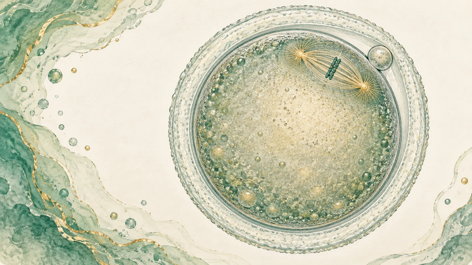
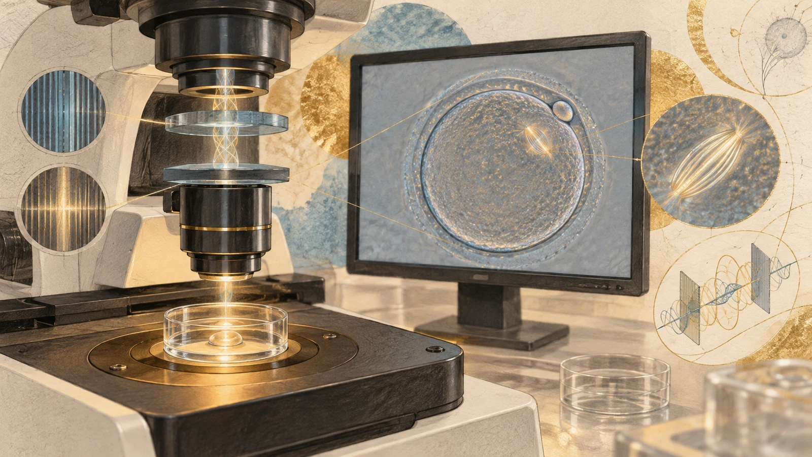
The traditional method to observe the spindle is to fix the oocytes and do tubulin immunofluorescence staining or transmission electron microscopy. Fixation means that the oocytes can no longer be used for treatment, and this type of method cannot enter the clinic. Polarized light microscopy does not require fixation and staining. Keefe et al. stated in a methodology paper in Reproductive BioMedicine Online in 2003 that liquid crystal polarized light microscopy (LC-PolScope) uses illumination close to circular polarization, so that the measured birefringence value does not depend on the orientation of the specimen in the imaging plane, and is sensitive enough to display low-level birefringence of mammalian spindles; the system has completed safety verification on mammalian oocytes, including human oocytes used for ICSI, and the structural information provided is close to immunostaining. [2]
Imaging is performed on a heated stage, typically maintained at 37°C. The entire observation process is completed within seconds, and the oocytes then proceed to ICSI as usual.
Visible proportions of the spindle under polarized light
Published polarized light visibility figures range from 78% to 86%. The age of the population, observation time and equipment in each study are different, and the numbers cannot be directly converted.
In a comparative observational study published by Fang et al. in the Journal of Assisted Reproduction and Genetics in 2007, 134 in vivo mature oocytes from 15 patients with an average age of 31.6±2.97 years, the spindle visibility rate under polarized light was 83.6%; 105 in vitro mature oocytes from 11 polycystic ovary syndrome patients with an average age of 31.09±1.13 years, the visibility rate was 77.1%. [3]
In the freeze–thaw cohort reported by Konc et al. in The Scientific World Journal in 2012, each oocyte underwent polarized light examination once before freezing and after recovery culture, and 221 spindles were detected out of 259 oocytes (85.3%). [4]
A retrospective study by Kratochvilova et al. published in Reproduction & Fertility in 2026 included 209 cycles in an assisted reproductive center in the Czech Republic from 2018 to 2024. The average age of women was 38.46±3.86 years old, of which 85% were over 35 years old. Of 1,879 oocytes first examined by polarized light 36 to 39 hours after triggering, 1,480 (78.8%) had visible spindles, 277 (14.75%) had no detectable spindles, and 122 (6.5%) showed the presence of connecting bridges between spindle microtubules and the first polar body. [14]
Another set of numbers that is often mixed up comes from fixation stain studies. Coticchio et al. reported in Human Reproduction in 2006 that 76 (73.1%) of 104 fresh, unfrozen MII oocytes showed normal equatorial arrangement of bipolar spindle-associated chromosomes under a confocal microscope. [5] This ratio measures whether the structure of the spindle is normal, and is not the same indicator as whether polarized light can detect birefringence signals.
Three situations corresponding to the invisible spindle
- The oocyte has not yet completed nuclear maturation. In oocytes that are in telophase I or are transitioning from meiosis I to meiosis II, the spindle is disintegrating or has not yet reassembled, and there is no stable microtubule arrangement to detect. Kratochvilova et al. combined this type of oocytes with the "connecting bridge" morphology and classified them into the immature group. [14]
- Microtubules depolymerize due to temperature drop. In an experiment by Yang et al. in the Journal of Experimental & Clinical Assisted Reproduction in 2010, 16 fresh MII oocytes with visible spindles at 37°C lost spindle signals after cooling to 20°C without cryoprotectant; the same batch of oocytes were treated with 1.5 M propylene glycol, 1.5 M ethylene glycol, 1.5 M dimethyl sulfoxide or dimethyl sulfoxide at 37°C. After equilibration with 10 μM paclitaxel and then cooling, the spindle is still visible at 20°C, 10°C and even 0°C, and some parts become clearer. The authors therefore recommend that the equilibrium temperature for oocyte freezing should not be lower than 33°C. [9]
Temperature-induced depolymerization is reversible upon rewarming. A review by Chen and Yang in the Taiwanese Journal of Obstetrics & Gynecology in 2009 pointed out that microtubules depolymerize under thermal changes and repolymerize after recovery in culture; spindle recovery after vitrification is faster than slow freezing, and the recovery degrees of the two methods are similar after 3 hours of incubation. [10]
- Abnormal spindle structure. Oocytes with disordered microtubule arrangement, multipolar spindles or chromosomes not aligned on the equatorial plane will have weak or shapeless birefringence signals. The prospective cohort published by Tilia et al. in Fertility and Sterility in 2020 divided the spindle morphology during ICSI into five categories: normal, malformed, translucent, invisible, and terminal. A total of 2,056 spindles were classified. "Invisible" was juxtaposed with "malformed" in this classification system and belonged to a group with poor outcomes. [11]
These three situations are indistinguishable in a single static observation. Kratochvilova et al.'s approach was to repeat observations about one hour later on oocytes that did not show spindles or connecting bridges during the first examination, and the final evaluation was scheduled about 20 minutes before ICSI. Repeated observations revealed changes in spindle signaling, but the reappearance of the signal did not alone distinguish maturation progression from microtubule reorganization after rewarming.
The positional relationship between the first polar body and the spindle
The conventional procedure for ICSI is to place the first polar body at the 12 or 6 o'clock position and insert the needle from the 3 o'clock position, provided that the spindle is immediately adjacent to the first polar body. This approximate positioning does not guarantee that the needle avoids the spindle.
Hardarson et al. recorded in Human Reproduction in 2000 the actual spatial relationship between the first polar body and the metaphase II spindle, expressed as the angle formed by the center of the oocyte, the spindle and the first polar body (the abstract of the article did not specify the imaging method). The average deviation of the spindle relative to the first polar body position of 54 oocytes matured in vivo was 41.7°, and that of 43 oocytes matured in vitro was 26.6°. The difference between the two groups was statistically significant (P=0.005). Most of the spindles in both groups were located in the same hemisphere as the first polar body. [6]
The same comparison was repeated in the Journal of First Military Medical University in 2004. Zhu et al. used LC-PolScope to observe that the angle difference between oocytes matured in vivo and matured in vitro was equally significant (P=0.006), and they concluded that the first polar body cannot predict the exact position of the metaphase II spindle. [7]
Maternal age is related to spindle position. In a retrospective analysis by Inoue et al. in Reproductive Medicine and Biology in 2024, the spindle was defined as 0° just below the first polar body. Multiple classification logistic regression showed that in older women, the odds ratios of spindles located at 30°<θ≤60°, 60°<θ≤90°, and 90°<θ≤180° relative to θ=0° were 1.020, 1.030, respectively. 1.060. [12]
Spindle status and fertilization rate, embryonic development and euploidy
Fertilization rate
In the 2007 study by Fang et al., the fertilization rate of oocytes with the spindle located directly below or immediately adjacent to the first polar body was 93.3%, which was significantly higher than that of the other groups; the fertilization rate of oocytes with no spindle detected decreased significantly in both in vivo and in vitro maturation categories. [3] In a retrospective analysis by Inoue et al. in 2024, under the four ovarian stimulation protocols of long regimen, short regimen, antagonist regimen and high progesterone ovarian stimulation (PPOS), the normal fertilization rate of oocytes with visible spindles was significantly higher than that of those without spindles; no difference was detected in the fertilization rate between each position interval of 0°≤θ≤180°. [12]
Blastocyst formation
The prospective cohort of Lee et al. in Frontiers in Endocrinology in 2026 included 581 MII oocytes from women aged 38 years and younger at a fertility center in Taiwan from August 2024 to August 2025. The available blastocyst formation rate was 46.1% for oocytes with visible metaphase II spindles and 15.6% for those without visible spindles. Oocytes with high birefringence signals in both the spindle and zona pellucida had the highest blastocyst rate of 53.7%. This study proposed the spindle-to-oocyte overall phase delay ratio (SOLRR) as a quantitative index. In multivariate analysis, SOLRR and anti-Müllerian hormone were positive and negative predictors of available blastocyst formation, respectively (adjusted odds ratios 1.148 and 0.939). The area under the receiver operating characteristic curve when SOLRR was used alone was at a moderate level. [13]
Euploidy rate
In the prospective cohort of Tilia et al. in 2020, normal spindle shape was positively correlated with blastocyst euploidy, and this association remained true after adjusting for maternal age, blastocyst quality, and developmental stage. [11] This suggests that the information carried by spindle morphology does not completely overlap with the information provided by age.
In the 2026 study by Kratochvilova et al., 462 (24.5%) of 1,879 oocytes developed into embryos and underwent preimplantation aneuploidy testing (PGT-A). 182 of 434 embryos from oocytes with visible spindles were euploid (42%); 4 of 28 embryos from oocytes with no visible spindle or junctional bridges were euploid (14%). The latter group has a denominator of 28 and estimates have limited accuracy. This study was a single-center retrospective design, and PGT-A was not performed on all embryos, but was performed selectively on high-risk patients, resulting in selection bias. [14]
"Extended culture" consists of two different operations
The timing studies address two different intervals.
The first type of extension is the interval between triggering and egg retrieval, as oocytes continue to mature within the follicle. This question has been studied in randomized controlled trials and systematic reviews. A meta-analysis by Wang et al. in the Journal of Assisted Reproduction and Genetics in 2011 included 5 randomized controlled trials with a total of 895 participants and reported that the proportion of mature oocytes in the long interval group was higher than that in the short interval group; the fertilization rate (RR 0.99, 95% CI 0.94–1.04), implantation rate (RR 0.91, 95% CI 0.40–2.04) and pregnancy rate (RR 0.79, 95% CI 0.58–1.08) no statistically significant difference was detected. [15]
A 2015 randomized controlled trial by Bosdou et al in Reproductive BioMedicine Online randomly assigned 156 normally ovulating women to have their eggs retrieved 36 or 38 hours after the trigger. The between-group differences were +1.2% in oocyte retrieval rate (95% CI −4.5 to +12.1 for median difference), median number of eggs retrieved 5.5 vs. 6.0, median fertilization rate 57.7% vs. 50.0%, and live birth rate 20.5% vs. 16.7% (rate difference +3.8%, 95% CI −8.5 to +16.1). No differences in outcomes were detected by extending the interval from 36 to 38 hours in this trial's population and endpoints. [16]
The idea of selective extension on an individual basis appeared in earlier randomized studies. Of 2,650 ICSI cycles, 72 cycles had 47% or more immature oocytes (Raziel et al., Fertility and Sterility 2006). These patients were assigned to inject the trigger drug at the same interval or 3 to 4 hours earlier in the next cycle, and the actual achieved trigger-to-ovum retrieval interval was 38.6 ± 1.2 hours versus 35.3 ± 0.7 hours, with no premature ovulation occurring. This design determines whether to extend based on the maturity of the previous cycle, and is suitable for patients with a high proportion of immature eggs in the past. [17]
Cohort data adjusting fertilization timing by spindle status
The operation procedure of Kratochvilova et al. is: complete the first polarized light examination 36 to 39 hours after the trigger; oocytes with no spindle or connecting bridge are reexamined about one hour later; if the cycle contains these two types of oocytes, the fertilization time of the cycle is postponed; the interval from the trigger to ICSI is controlled between 36 and 44 hours, and not more than 44 hours. The reason given by the author is the risk of oocyte aging. [14]
After this process, the proportion of oocytes with visible spindles increased from 78.8% (1,480/1,879) in the first examination to 92.2% (1,733/1,879) in the final examination, those without spindles dropped from 14.75% (277) to 6.2% (116), and those with connecting bridges fell from 6.5% (122) to 1.6% (30). The change in signal may reflect maturation or microtubule recovery after warming; these data alone cannot separate the two.
Euploidy rates grouped by trigger to ICSI interval: 76 of 230 embryos (33%) in the 36 to 39 hour group, 110 of 232 embryos in the 40 to 44 hour group (47%), P=0.039557.
The average age of women in this cohort was 38.46 years, with 85% over 35 years old, and the results correspond to an older age group.
Applications of spindle observation in other scenarios
Early rescue ICSI after conventional IVF fertilization failure
Gao et al's 2025 retrospective cohort in the Journal of Assisted Reproduction and Genetics included 332 early salvage ICSI cycles and were divided into two groups according to whether polarized light spindle observation was performed before salvage. The normal fertilization rate in the observation group was 82.55% and that in the control group was 71.49% (P<0.01); the three pronucleus rates were 3.17% and 8.89% respectively (P<0.01). In the subgroup over 35 years old, the normal cleavage rate was 38.60% in the observation group and 22.47% in the control group (P<0.05). No statistically significant differences were detected in embryonic development and implantation potential between the two groups. [18]
Timing of oocyte freezing
In the case reported by Mašata et al. in "Česká gynekologie" in 2024, the oocytes of a 38-year-old woman were classified according to the polarization results. The oocytes whose spindle and first polar body were not more than 30° or whose spindle was unclear were cultured for another 4 hours after evaluation (8 hours after egg retrieval) before being vitrified. After thawing, they were matured and fertilized, and finally delivered at term. Case reports do not provide comparisons, only descriptions of how things work. [20]
Grading of spindle morphology
Buderatska et al. proposed the MOL score in the Journal of Assisted Reproduction and Genetics in 2026, using three-digit codes to record spindle morphology (barrel-shaped, changed, enlarged, unclearly displayed, missing), orientation and angular displacement relative to the first polar body. This study simultaneously documents the effects of cryo-thaw on three distributions, reporting that specific MOL combinations are associated with fertilization and euploid blastocyst formation. [19]
Automatic quantification of the angle between spindle and polar body
A 2026 preprint used a U-Net-based deep learning model to automatically measure the spindle-polar body angle of MII oocytes imaged approximately 40 hours after 81 triggers, reporting an euploidy rate of 72.2% for oocyte-derived embryos falling within the 8° to 32° interval and 31.1% outside the interval (P=0.000236). The odds ratio for patient cluster logistic regression was 5.76 (95% CI 2.03–16.31). This article is a proof-of-concept study with 81 samples and has not yet been peer-reviewed. [21]
Current state of evidence
The feasibility and safety of polarized-light spindle imaging have been studied at the methodological level (Keefe et al. 2003; Zhu et al. 2004). Its association with outcomes has been replicated in multiple independent cohorts at the level of fertilization rates and embryonic development (Fang et al. 2007; Tilia et al. 2020; Inoue et al. 2024; Lee et al. 2026; Kratochvilova et al. 2026).
The evidence at the clinical endpoint level is much weaker. In the retrospective analysis by Inoue et al. in 2024, no differences were detected in the pregnancy, live birth or ongoing pregnancy rates, and miscarriage rate between visible and invisible spindles, or between various positions of the spindle. In the study by Gao et al. in 2025, the differences between groups in fertilization rate and cleavage rate reached statistical significance, and no differences were detected in embryonic development and implantation potential. The studies reviewed here do not include randomized controlled trials assessing live birth after spindle-guided timing.

For older patients, what can be read from the existing data is that some telophase I oocytes may be counted as MII based on the first polar body alone; polarized light observation can identify a part of the oocytes that have not completed nuclear maturation; in single-center retrospective data, after postponing fertilization of these oocytes, the proportion of spindles can be seen to increase, and the euploidy rate of embryos in the corresponding cycle is higher than that in cycles fertilized at regular intervals. In that centre, ICSI was performed no later than 44 hours after the trigger.
Documentation and evidence table
Design, sample and main results of 21 articles
| Literature | Design and Sample | Primary Endpoints and Results |
|---|---|---|
| [1] ESHRE/ALPHA. Istanbul consensus update. Hum Reprod 2025;40(6):989 | Society consensus document | Oocytes with first polar body may be in telophase I; only observation of metaphase II spindles confirms nuclear maturation; evidence is insufficient to recommend routine use |
| [2] Keefe D, Liu L, Wang W, Silva C. Reprod Biomed Online 2003;7(1):24–29. PMID 12930570 | Methodology Paper | Principle of LC-PolScope; circularly polarized illumination enables measurements independent of in-plane orientation; safety verified in mammalian oocytes containing oocytes from ICSI patients |
| [3] Fang C, Tang M, Li T, et al. J Assist Reprod Genet 2007;24(11):547–551. PMID 17899355 | Comparative observational study; in vivo maturation 134 pieces/15 cases (31.6±2.97 years old), in vitro maturation 105 pieces/11 cases PCOS (31.09±1.13 years old) | The spindle visibility rate was 83.6% vs. 77.1%; the fertilization rate of those with the spindle located directly below or immediately adjacent to the first polar body was 93.3%, which was significantly higher than other groups |
| [4] Konc J, Kanyo K, Kriston R, et al. ScientificWorldJournal 2012;2012:785421. PMID 22629197 | Freeze-resuscitation cohort, 259 pieces; checked once before freezing and after recovery culture | Spindles detected by polarized light 221/259 (85.3%); recovery survival rate 81.1% |
| [5] Coticchio G, De Santis L, Rossi G, et al. Hum Reprod 2006;21(7):1771–1776. PMID 16549422 | Confocal microscopy comparison, fresh set of 104 pieces | Normal bipolar spindle with equatorial aligners of chromosomes in fresh MII oocytes 76/104 (73.1%); slow-frozen low sucrose group 50.8% (61 pieces), 0.3 mol/l sucrose group 69.7% (152 pieces) |
| [6] Hardarson T, Lundin K, Hamberger L. Hum Reprod 2000;15(6):1372–1376. PMID 10831572 | Angle measurement; 54 pieces matured in vivo, 43 matured in vitro | The average deviation of the spindle relative to the first polar body is 41.7° with 26.6° (P=0.005); most located in the same hemisphere |
| [7] Zhu L, Kong LH, Li H, et al. "Journal of First Military Medical University" 2004;24(7):809–811. PMID 15257910 | LC-PolScope observation | The angle of mature oocytes in vivo and in vitro is significantly different (P=0.006); the first polar body cannot predict the exact position of the spindle |
| [8] Rienzi L, Ubaldi F, Iacobelli M, et al. Reprod Biomed Online 2005;10(2):192–198. PMID 15823222 | Discussion paper | The spindle of mature oocytes in vitro is aligned with the first polar body, and the misalignment of mature oocytes in vivo is attributed to the denudation operation; consistent with the literature [6] The data are inconsistent with the interpretation |
| [9] Yang D, Winslow KL, Nguyen K, et al. J Exp Clin Assist Reprod 2010;7:4. PMID 20941373 | Experimental study, 26 fresh MII oocytes | When cooled to 20℃ without cryoprotectant, the spindle signal disappears; after adding PROH/EG/DMSO/paclitaxel 20℃, 10℃, 0℃ are still visible; recommended equilibrium temperature ≥33℃ |
| [10] Chen SU, Yang YS. Taiwan J Obstet Gynecol 2009;48(1):15–22. PMID 19346187 | Review | Microtubules depolymerize due to thermal changes and then repolymerize after incubation; recovery after vitrification is faster than slow freezing, and is similar after 3 hours of incubation |
| [11] Tilia L, Chapman M, Kilani S, Cooke S, Venetis C. Fertil Steril 2020;113(1):105–113.e1. PMID 31739977 | Prospective cohort, 2,056 spindle morphologies | Spindle morphology and fertilization, 3 There is a correlation between day cleavage and the formation of high-quality blastocysts; normal morphology is positively correlated with blastocyst euploidy, and this is still true after correcting for the woman's age, blastocyst quality and developmental stage |
| [12] Inoue T, Matsuo Y, Taguchi S, et al. Reprod Med Biol 2024;23(1):e12601. PMID 39677331 | Retrospective analysis, four ovarian stimulation regimens | The normal fertilization rate of those with visible spindles was significantly higher; no differences were detected in pregnancy rate, live birth/continuous pregnancy rate, and miscarriage rate between visibility and position; increasing age is associated with spindle deviation from the first polar body (OR 1.020/1.030/1.060) |
| [13] Lee CI, Chen HH, Lo WC, et al. Front Endocrinol 2026;17:1803476. PMID 42165024 | Prospective cohort, 581 MII oocytes, female ≤38 years old | Available blastocyst rate in the visible spindle group was 46.1%, in those without 15.6%; those with high spindle and zona pellucida birefringence were 53.7%; SOLRR and AMH were significant predictors in multivariate analysis (aOR 1.148 and 0.939) |
| [14] Kratochvilova I, Tepla O, Pšeničková A, et al. Reprod Fertil 2026;7(3):RAF260016. PMID 42496869 | Single center retrospective, 209 cycles, 1,879 oocytes, female 38.46±3.86 years old, 85% >35 years old | Visible spindle proportion 78.8% → 92.2%; euploidy 182/434 (42%) with visible spindles versus 4/28 (14%) without visible spindles or with connecting bridges; 76/230 (33%) at 36–39 h versus 110/232 (47%) at 40–44 h (P=0.039557). Timing was assigned by the first assessment, not randomized; PGT-A was selective. |
| [15] Wang W, Zhang XH, Wang WH, et al. J Assist Reprod Genet 2011;28(10):901–910. PMID 21792666 | Meta-analysis, 5 RCTs, 895 people | A longer interval increased the proportion of mature oocytes. No statistically significant differences were detected for fertilization (RR 0.99, 95% CI 0.94–1.04), implantation (RR 0.91, 95% CI 0.40–2.04) or pregnancy (RR 0.79, 95% CI 0.58–1.08). |
| [16] Bosdou JK, Kolibianakis EM, Venetis CA, et al. Reprod Biomed Online 2015;31(5):625–632. PMID 26387934 | Randomized controlled trial, 156 people, 36 h vs 38 h | Difference in oocyte retrieval rate +1.2% (median difference 95% CI −4.5 to +12.1); live birth rate 20.5% vs 16.7% (rate difference +3.8%, 95% CI −8.5 to +16.1) |
| [17] Raziel A, Schachter M, Strassburger D, et al. Fertil Steril 2006;86(3):583–587. PMID 16828475 | Randomized study, 72 previous cycle immature eggs ≥47% of cycles | Achievement interval 38.6±1.2 h vs 35.3±0.7 h; no premature ovulation occurred |
| [18] Gao Y, Zhu J, Xiong S, et al. J Assist Reprod Genet 2025;42(5):1625–1633. PMID 40195249 | Retrospective cohort, 332 early salvage ICSI cycles | Normal fertilization rate 82.55% vs 71.49% (P<0.01); 3PN rate 3.17% vs 8.89% (P<0.01); the normal cleavage rate in the subgroup >35 years old was 38.60% vs 22.47% (P<0.05); no difference was detected in embryonic development and implantation potential |
| [19] Buderatska N, Gontar J, Yurchuk A, et al. J Assist Reprod Genet 2026;43(1):229–243. PMID 41191210 | Research on the establishment of MOL scoring system | Three-dimensional coding of morphology, orientation, and positioning; cryogenic recovery changes the distribution of three items; specific combinations are related to fertilization and euploid blastocyst formation |
| [20] Mašata J, Teplá O, Jirsová S, et al. "Česká gynekologie" 2024;89(6):475–478. PMID 39800544 | Case report and literature review, 38-year-old woman | Oocytes with an angle ≤30° or unclear spindle signal underwent four additional hours of culture after assessment, then vitrification eight hours after retrieval; fertilization and transfer after warming led to a term birth in this case. |
| [21] Automatic quantification of spindle-polar body angle. Preprint, DOI 10.21203/rs.3.rs-10856040/v1 | Proof-of-concept, 81 MII oocytes imaged ~40 h after triggering | 8°–32° interval euploidy rate 72.2% vs 31.1% outside the interval (P=0.000236), OR 5.76 (95% CI 2.03–16.31); not peer-reviewed |
Table note: The percentages in the literature [3][4][5] respectively correspond to the polarized light visible rate and the structural normality rate under confocal. The two types of indicators are not directly comparable. Document [15][16][17] prolongs the interval from trigger to egg retrieval, and document [14] prolongs the interval from trigger to ICSI, the latter including the in vitro culture time after egg retrieval. The evidence search ended on September 22, 2026, and the author, journal, volume, and page number were checked item by item by Europe PMC; no registered systematic review was conducted.
Medical notice
This article is medical popular science content, compiled based on published literature. It does not replace face-to-face consultation, does not constitute diagnosis and treatment recommendations, and does not target any specific patient's treatment plan. The spindle observation, fertilization timing adjustment and laboratory procedures involved in this article are determined by each reproductive center according to its own conditions and local regulations.
All quantitative results in this article come from the original abstracts or full texts of the literature listed at the end of the article. The design and sample size are noted along with the results. Single-center retrospective data and randomized controlled trials have different strengths of evidence and are noted separately in the table.
The article includes five medical infographics and three AI-generated concept illustrations. The web cover uses one of the concept illustrations. None depicts real microscopy or clinical data.
Interpret laboratory markers in context
FS can help organize retrieval, ICSI, embryo culture and testing records. The treating team must decide whether any timing change is appropriate for a specific cycle.
Consensus and original studies
Observational differences in euploidy do not establish an individual live-birth benefit from delaying ICSI.
- ESHRE/ALPHA Istanbul consensus update, Human Reproduction 2025
- Research reference [2], PubMed 12930570
- Research reference [3], PubMed 17899355
- Research reference [4], PubMed 22629197
- Research reference [5], PubMed 16549422
- Research reference [6], PubMed 10831572
- Research reference [7], PubMed 15257910
- Research reference [8], PubMed 15823222
- Research reference [9], PubMed 20941373
- Research reference [10], PubMed 19346187
- Research reference [11], PubMed 31739977
- Research reference [12], PubMed 39677331
- Research reference [13], PubMed 42165024
- Research reference [14], PubMed 42496869
- Research reference [15], PubMed 21792666
- Research reference [16], PubMed 26387934
- Research reference [17], PubMed 16828475
- Research reference [18], PubMed 40195249
- Research reference [19], PubMed 41191210
- Research reference [20], PubMed 39800544
- Research reference [21], preprint; not peer reviewed
For public medical education only; not individual medical advice.