

RCOG 2024 · Nature 2024 · Nature Genetics 2026 · ASRM 2018/2022/2024/2026 · ACOG
Weeks of severe nausea and vomiting, rapid weight loss and repeated intravenous fluids should first be assessed as hyperemesis gravidarum and its complications. A gestational carrier is not genetically related to the embryo, but that is not equivalent to organ-transplant rejection. A PGT-A euploid result does not predict vomiting, placental function or every pregnancy risk.
When severe, persistent vomiting prevents normal eating and drinking and is accompanied by substantial weight loss, dehydration, electrolyte disturbance or a need for intravenous fluids, the first clinical consideration is hyperemesis gravidarum (HG). This pregnancy complication can cause systemic harm and should not be dismissed as ordinary morning sickness.[1-3]
The absence of a genetic relationship between the gestational carrier and the embryo does not mean her immune system will reject the embryo as it might reject a transplanted kidney or liver. Healthy pregnancy establishes maternal-fetal immune tolerance through decidua, placental trophoblast, uterine natural killer cells, regulatory T cells and multiple local signals. Pregnancy is a distinct reproductive-immune state; the organ-transplant model cannot simply be applied to it.[6-8]
A PGT-A report of euploidy means that no clear chromosome-number abnormality was detected within the assay's target range in the biopsied sample. It may guide embryo selection, but it does not test susceptibility to HG and cannot guarantee implantation, an ongoing pregnancy, normal placental function or the absence of every genetic condition in a child.[9-10]
The strongest current biological explanation for severe pregnancy sickness is the interaction between a rapid rise in GDF15 produced by the fetal-placental unit and the pregnant person's sensitivity to GDF15. Multiple pregnancy, a prior history of HG and genetic susceptibility may increase risk. Ultrasound and laboratory testing are still needed to exclude multiple gestation, molar pregnancy, infection, thyroid disease and gastrointestinal disease.[1-5]
1 | When symptoms are no longer ordinary pregnancy sickness
Nausea, reduced appetite and occasional vomiting are common in early pregnancy. For most people symptoms begin in the first trimester and improve during the second. HG lies at the severe end of the spectrum: patients may be unable to take enough fluid or food for prolonged periods, their daily activity becomes markedly restricted, and dehydration, undernutrition, rapid weight loss and metabolic disturbance may follow.[1-3]
Older definitions often used weight loss above 5% of pre-pregnancy weight or urinary ketones. Modern guidance stresses a combined assessment: symptom severity and duration, actual intake, weight trend, vital signs, urine output, electrolytes, liver and kidney function, and impact on daily life. The 2024 RCOG guideline states that ketonuria alone does not measure dehydration and should not be the sole threshold for treatment.[1]
Table 1 | Distinguishing common pregnancy sickness from HG
| Domain | Common nausea and vomiting | Features suggesting HG |
|---|---|---|
| Food and fluids | Nausea is prominent, but small frequent intake is retained | Fluids are vomited soon after drinking; 24-hour intake is clearly inadequate |
| Weight | Stable or mildly reduced | Continues to fall over a short period |
| Vital signs | Normal | Postural dizziness, palpitations, weakness or syncope |
| Urine | Normal | Reduced output or very dark urine |
| Antiemetics | Oral medication is retained and works | Oral medication cannot be retained; another route is needed |
| Laboratory tests | No major abnormality | Low potassium or sodium, acid-base disturbance, abnormal liver tests or pre-renal injury |
| Level of care | Outpatient follow-up | Repeated emergency, day-unit or inpatient fluids with rapid relapse |
The following features go beyond mild pregnancy sickness:
- even water cannot be retained and is vomited shortly after drinking;
- 24-hour intake is clearly inadequate, with reduced or very dark urine;
- weight continues to fall over a short period;
- standing causes dizziness, palpitations, weakness or syncope;
- oral antiemetics cannot be retained and injections, intravenous medication or another route is required;
- blood tests show low potassium or sodium, acid-base disturbance, abnormal liver function or pre-renal injury;
- repeated emergency, day-unit or inpatient fluids are needed and symptoms quickly rebound when fluids stop.
A repeated need for intravenous fluids is itself an important severity signal. Treatment must do more than stop vomiting: it should correct volume depletion, electrolyte and vitamin deficiencies, restore sustainable intake, and prevent uncommon but serious complications such as thrombosis and Wernicke encephalopathy.[1-3]
2 | Why a gestational-carrier pregnancy usually does not cause transplant-style rejection
Although the gestational carrier and embryo are not genetically related, a normal pregnancy creates specialised immune tolerance at the uterine-placental interface. It is unlike organ transplantation: donor-recipient HLA matching is not required, and the pregnancy does not undergo the characteristic acute rejection seen in a transplanted organ.
The anatomy of the maternal-fetal interface explains why. After implantation, placental trophoblast cells—not fetal organs directly—form the principal contact with maternal tissue. Trophoblast has a specialised pattern of HLA expression, and immune cells in the decidua differ substantially from cells with similar names in peripheral blood. Their continuing signals regulate trophoblast invasion, spiral-artery remodelling, local inflammation and immune tolerance.[6-7]
Uterine natural killer cells are often called uNK or dNK cells. The word killer is easily misunderstood. In early-pregnancy decidua these cells mainly support vascular remodelling, trophoblast regulation and local homeostasis; their phenotype and function are not equivalent to peripheral-blood NK cells that kill infected or malignant cells. Regulatory T cells, macrophages, dendritic cells and multiple cytokines also support tolerance. The maternal immune system is not switched off. It protects against infection while applying fine, dynamic local regulation to placental development.[6-8]

Calling pregnancy a natural allograft can help frame research questions, but it easily produces clinical misunderstanding. Organ-transplant rejection typically involves systemic recognition of graft antigens, cytotoxic responses, antibody-mediated injury and progressive loss of graft function. Normal pregnancy has a unique placental structure, HLA expression, endocrine environment and local immune regulation. Both involve recognition of self and non-self, but their biology, clinical manifestations and management are not the same.
Table 2 | Organ-transplant rejection and immune tolerance in pregnancy
| Comparison | Solid-organ transplant | Pregnancy |
|---|---|---|
| Contact interface | Graft vasculature broadly contacts the recipient immune system | Mainly trophoblast and decidua; fetal organs are not directly exposed |
| HLA expression | Classical HLA is routinely expressed and matching is used | Trophoblast has specialised HLA expression; matching is not required |
| Local immune cells | Peripheral effector cells infiltrate and mediate cytotoxic injury | Decidual uNK and regulatory T cells mainly support vascular remodelling and tolerance |
| Typical injury | Acute or chronic rejection with progressive organ dysfunction | No equivalent pattern of acute rejection |
| Routine medication | Long-term immunosuppression | No preventive immunosuppression |
| Pre-treatment process | HLA and cross-matching of donor and recipient | Medical and infection screening, psychosocial assessment and informed consent |
Accordingly, gestational-carrier programmes do not routinely match the HLA of embryo and carrier, nor use transplant-level immunosuppression simply because they are not genetically related. ASRM guidance focuses on full medical and infectious-disease screening, psychosocial assessment, genetic-source screening, informed consent and continuous obstetric care; routine HLA matching is not a prerequisite for transfer.[12]

3 | Immune biology matters in pregnancy, but not every abnormality is rejection
Maternal-fetal tolerance does not mean immune factors are irrelevant to pregnancy complications. Trophoblast invasion, decidual immune cells, complement, coagulation and vascular remodelling have complex links; abnormalities may be associated with implantation failure, miscarriage, placental dysfunction or pre-eclampsia. The crucial distinction is between an immune mechanism existing and a routine clinical test being able to diagnose embryo rejection.
No routine clinical test can prove that a pregnant person is rejecting an embryo when implantation has occurred, ultrasound growth matches gestation and the principal problem is severe vomiting. Peripheral NK-cell count or activity does not directly represent uterine immunity. Th1/Th2 ratios, cytokine panels, uterine NK-cell counts and KIR/HLA combinations also lack sufficiently standardised measurement, reference ranges and reproducible treatment thresholds.
ASRM's 2018 IVF immunotherapy guideline found no reliable evidence that routine peri-implantation corticosteroids improve live birth in a general IVF population; evidence is also insufficient for routine intralipid or intravenous immunoglobulin use.[8] ASRM's 2026 recurrent pregnancy loss opinion further emphasises that even in recurrent loss, including recurrent euploid loss—a population of particular research interest—immune tests and treatments should remain mainly within research and are not recommended as routine care.[11]
This does not deny the medical importance of defined diseases such as antiphospholipid syndrome, systemic lupus erythematosus or autoimmune thyroid disease. When history or clinical criteria indicate them, assessment and treatment should follow the relevant guidelines. These conditions have diagnostic criteria and should not be collapsed into the vague label of a rejection-prone constitution.
4 | What evidence usually appears if implantation or pregnancy is failing
Failed implantation commonly appears as β-hCG that never reaches the positive threshold after transfer, or rises briefly and then falls. Once a clinical pregnancy is established, an abnormal course may involve an inappropriate β-hCG trend, delayed gestational sac or embryo development, loss of cardiac activity, vaginal bleeding, pain or miscarriage. Ultrasound and serial clinical data are more informative about pregnancy progression than a single symptom.
Severe vomiting alone proves neither that a pregnancy is healthy nor that an embryo is being rejected. Symptoms are influenced by GDF15, maternal sensitivity, placental mass, fetal number and individual susceptibility. A normally developing pregnancy may cause little nausea, while nausea can persist for a time in an abnormal pregnancy before hormone levels fall. Using symptom intensity to infer embryo quality, heartbeat or chromosome status can delay necessary ultrasound and laboratory assessment.
If ultrasound confirms an intrauterine pregnancy with an embryo and cardiac activity growing broadly in line with gestational age, persistent vomiting and dehydration fit HG more closely. If vaginal bleeding, progressive pain, abnormal ultrasound growth or an abnormal β-hCG trend is also present, pregnancy viability must be assessed separately rather than merging every problem into a rejection diagnosis.
5 | The strongest current mechanism for severe vomiting: GDF15 and maternal sensitivity
Growth differentiation factor 15 (GDF15) is a circulating protein that can activate a specific hindbrain receptor pathway and cause reduced appetite, nausea and vomiting. In pregnancy, the fetal-placental unit produces large amounts of GDF15, and maternal blood levels rise rapidly.
A 2024 Nature study found that most GDF15 in maternal circulation during pregnancy comes from the fetal-placental unit. GDF15 was higher in patients with HG, while maternal sensitivity to the pathway also influenced severity. People with chronically lower GDF15 before pregnancy may respond more strongly when fetal GDF15 rises quickly. This model connects fetal genetics, placental secretion and maternal susceptibility in one pathway.[4]
A large multi-ancestry genome-wide association study published in 2026 included 10,974 cases of severe pregnancy nausea and vomiting and 461,461 controls. It identified ten associated loci across European, Asian, African and Latino populations. The GDF15/GFRAL pathway remained central, with additional pathways involving appetite, insulin signalling, the progesterone receptor and brain plasticity. HG therefore has a defined biological basis and is not explained by one hormone or a psychological factor alone.[5]
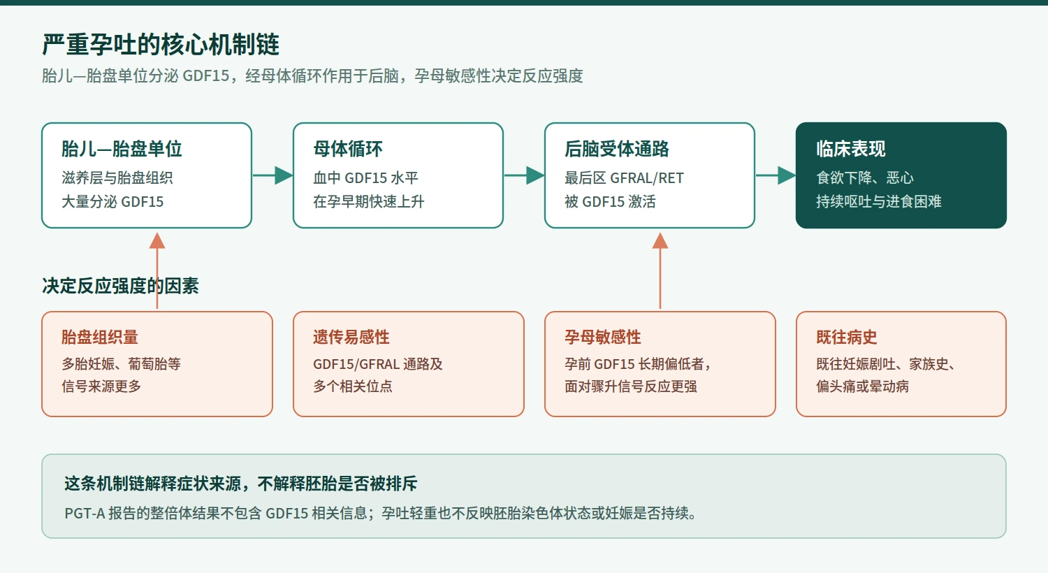

hCG, oestrogen, progesterone, the thyroid axis and gastrointestinal motility may contribute to or modulate symptoms, but this does not support the rule that higher hCG always means worse vomiting. Multiple pregnancy and molar pregnancy, which involve more placental tissue or an abnormal hormonal environment, are associated with higher risk and should be assessed by ultrasound. Luteal-support medication can cause gastrointestinal symptoms, but rapid weight loss, dehydration and inpatient fluids should not be attributed solely to oestrogen or progesterone—and medication should not be stopped without clinical advice.
6 | What a PGT-A euploid result actually means
PGT-A is preimplantation genetic testing for aneuploidy. A small number of cells are generally taken at the blastocyst stage from the trophectoderm, which will mainly contribute to the placenta, and chromosome copy number is assessed to guide embryo selection. Euploid means that no clear chromosome-number abnormality was detected in that sample within the laboratory's technical scope and interpretation rules.[9]
The information has practical value. In some patients PGT-A can reduce the chance that a clearly aneuploid embryo is prioritised, help rank several embryos and support single-embryo transfer. It may offer value especially to older patients who have several blastocysts. At the same time, ASRM's 2024 opinion states that a cumulative live-birth benefit from routine PGT-A for every IVF patient has not been consistently demonstrated; age, ovarian reserve, embryo number and laboratory conditions materially affect its value.[9]
Table 3 | Information boundaries of a euploid PGT-A report
| Domain | PGT-A can provide | PGT-A cannot provide |
|---|---|---|
| Sample | A few trophectoderm cells from a blastocyst | Every embryo cell, including the inner cell mass |
| Target | Chromosome-number information within the assay range | All monogenic, epigenetic and structural-development disorders |
| Nature of result | A laboratory-specific technical interpretation | An absolute result unaffected by biopsy, amplification or platform resolution |
| Clinical use | Embryo ranking and support for single-embryo transfer | A guarantee of implantation, ongoing pregnancy or live birth |
| Pregnancy risks | Some information on chromosome-number risk | Placental function, uterine environment, maternal disease, infection or cord factors |
| Vomiting | None | Prediction of GDF15 sensitivity or HG |
| Prenatal care | One input to genetic counselling | A replacement for prenatal screening or diagnosis |
A euploid PGT-A result does not mean the embryo is normal in every respect. Reasons include:
- only a limited number of trophectoderm cells are tested, not every embryo cell;
- cell-to-cell variation or mosaicism may mean the sample does not always match the inner cell mass;
- the test primarily targets chromosome copy number and does not cover every monogenic, epigenetic or structural-development condition, or problems arising later in pregnancy;
- results depend on biopsy, DNA amplification, platform resolution, quality control and laboratory interpretation;
- placental development, the uterine environment, maternal disease, infection, cord factors and other obstetric issues fall outside PGT-A's direct scope.

ACOG states that a negative preimplantation genetic-test result cannot guarantee a newborn without genetic abnormality; false-positive and false-negative results can occur. After transfer of a PGT-A euploid embryo, prenatal screening and diagnostic options should still be discussed according to the pregnant person's preferences and local guidance.[10]
PGT-A does not test GDF15 sensitivity and does not predict HG. There is therefore no contradiction in asking why severe vomiting occurred after transfer of a euploid embryo. One result describes chromosome copy number in an embryo-biopsy sample; the other condition is a clinical syndrome produced by placental signalling, maternal physiology and the pregnancy environment.
7 | Does gestational-carrier status increase the risk of HG?
Gestational-carrier status alone does not support the inference that severe vomiting is caused by immune rejection. Better-established HG risk factors include prior HG, family history, multiple pregnancy, migraine and motion sickness.[2-3]
Several variables need extra attention in assisted reproduction. First, transfer of more than one embryo, or splitting of a single embryo, can produce a multiple pregnancy; multiple gestation is associated with more severe nausea and vomiting. Second, frozen-embryo transfer cycles often use oestrogen and progesterone for endometrial preparation and luteal support. Their gastrointestinal effects may overlap with early-pregnancy symptoms, but severe dehydration and rapid weight loss still require HG assessment. Third, more participants, institutions and intended parents are involved in a gestational-carrier programme. Delayed communication can allow steadily worsening symptoms to be treated as something the carrier must simply endure.
The gestational carrier needs clear authority and an escalation pathway for medical care. She has the right to report symptoms directly, receive an independent medical assessment, understand medication and participate in treatment decisions. No contract, travel plan or programme schedule should override urgent medical need. The clinical team should retain complete transfer, medication, ultrasound, weight, fluid and antiemetic records for continuity of care.[12]
8 | Other diseases that severe vomiting must exclude
HG is a clinical diagnosis made when another disorder does not better explain the symptoms. Typical nausea and vomiting early in pregnancy without pain, fever or focal signs is most often pregnancy-related. Very severe symptoms, atypical timing or associated signs require a broader differential diagnosis.[1-3]
Obstetric causes include multiple pregnancy, molar pregnancy, ectopic pregnancy and ovarian torsion. Gastrointestinal causes include gastroenteritis, hepatitis, gallbladder disease, pancreatitis, peptic ulcer and bowel obstruction. Urinary infection and pyelonephritis must be considered. Endocrine and metabolic causes include thyroid disease, diabetic ketoacidosis, adrenal insufficiency and major electrolyte disturbance. Neurological disease, migraine and medication adverse effects may also cause persistent vomiting.
Table 4 | Differential diagnosis checklist
| System | Conditions to exclude | Clues |
|---|---|---|
| Obstetric | Multiple or molar pregnancy, ectopic pregnancy, ovarian torsion | Abnormal number or anatomy on ultrasound, bleeding, unilateral pain |
| Gastrointestinal | Gastroenteritis, hepatitis, gallbladder disease, pancreatitis, ulcer, obstruction | Pain, diarrhoea, jaundice, haematemesis or melaena, absent flatus or stool |
| Urinary | UTI, pyelonephritis | Dysuria, fever, flank tenderness |
| Endocrine/metabolic | Thyroid disease, diabetic ketoacidosis, adrenal insufficiency | Palpitations, tremor, abnormal glucose, deep breathing, drowsiness |
| Neurological | Intracranial disorder, migraine | Severe headache, neck stiffness, altered consciousness, focal signs |
| Medication | Adverse effect from another drug | Timing matches a newly started medication |
The following features are especially unsuitable for simple classification as HG:
- marked or progressively worsening abdominal pain or rebound tenderness;
- fever, rigors or dysuria;
- severe headache, neck stiffness, altered consciousness or focal neurological signs;
- vomiting that first begins after 11 weeks' gestation;
- haematemesis, melaena, jaundice or persistent chest pain;
- markedly abnormal glucose with deep breathing or drowsiness suggesting metabolic crisis;
- failure to confirm an intrauterine pregnancy on ultrasound, or bleeding with unilateral pain.
Ultrasound has two jobs here: to confirm gestational age, embryo development and cardiac activity, and to establish fetal number while excluding molar pregnancy and other abnormalities. It supplies direct structural evidence. The severity of vomiting cannot perform that function.
9 | Objective measurements for specialist assessment
Assessment of a patient who needs intravenous fluids cannot stop at today's number of vomiting episodes. A continuous, comparable record should include:
- gestational week at onset, daily duration of nausea, number of vomiting episodes, excessive salivation and triggers;
- actual fluid and food intake over 24 hours, and whether oral medicines are retained;
- pre-pregnancy weight, current weight and weekly trend—not a single measurement;
- blood pressure, pulse, temperature, respiratory rate and symptoms on postural change;
- urine volume, colour and frequency;
- full blood count, electrolytes, urea, creatinine, glucose and liver tests, with magnesium, phosphate, thyroid, infection and pancreatic or biliary markers when indicated;
- ultrasound findings including fetal number, gestational age, cardiac activity and placental or trophoblastic abnormalities;
- name, dose, route, effect and adverse effects of antiemetics used;
- history of HG, migraine, motion sickness, gastrointestinal or thyroid disease, and mental-health history;
- thrombosis risk, mobility, nutritional risk and emotional state.
HG can cause transient thyroid-test changes, particularly lower TSH and higher free thyroxine. Without clinical evidence of Graves disease, and when values improve with fluids and antiemetics, one abnormal panel should not automatically be labelled primary hyperthyroidism. Further testing and treatment depend on thyroid signs, antibodies, symptoms and trends.[1]
10 | Treatment priorities: fluids, antiemetics, vitamins and complication prevention
HG treatment should be stratified by severity. A patient who can drink small amounts, has stable vital signs and no major laboratory abnormality may be managed with outpatient antiemetics, dietary adjustment and close follow-up. Inability to maintain intake, failed oral medication, clear dehydration, electrolyte disturbance or rapid weight loss requires day-unit fluids or admission.[1-3]
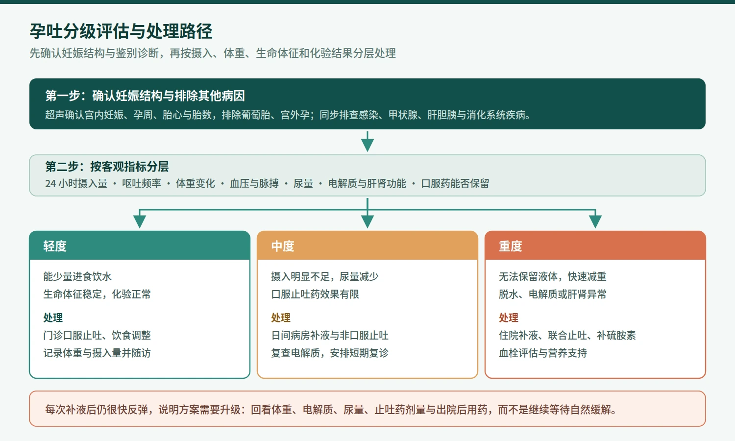
Intravenous fluid composition should follow electrolyte results. RCOG recommends 0.9% sodium chloride as the principal fluid base, with potassium added according to monitoring. Prolonged vomiting or markedly reduced intake requires thiamine, or vitamin B1. Thiamine-deficiency risk should be addressed before glucose-containing fluid or parenteral nutrition to prevent Wernicke encephalopathy.[1]

Antiemetic treatment usually begins with medicines that have pregnancy safety data, with different mechanisms combined after clinical assessment if one medicine is insufficient. Choice depends on gestation, severity, previous response, electrocardiographic risk, extrapyramidal adverse effects, constipation and sedation. Concern about medication should not leave a patient unable to eat for prolonged periods; untreated dehydration and malnutrition also create maternal and fetal risk. Prescribing must remain individualised by the responsible obstetric or maternal-fetal medicine team.[1-3]
Bed rest, dehydration and pregnancy all increase venous-thromboembolism risk. Inpatients need thrombosis-risk assessment and mechanical or pharmacological prevention when indicated. Continuing weight loss also requires dietetic input for energy, protein and micronutrient support. Rare cases unresponsive to structured fluids, combined antiemetics and nutrition may need higher-level treatment under specialist risk-benefit review.
Maintenance fluids should not become repeated bags of the same fluid without reassessment. Every relapse should trigger review of weight, electrolytes, urine output, antiemetics, thiamine and the post-discharge regimen. Improvement only during the infusion means the plan needs escalation, not continued waiting for spontaneous relief.
11 | Statements that need correction
1. The more severe the vomiting, the better the embryo
There is no such diagnostic relationship. Nausea and vomiting are associated with pregnancy hormones and GDF15, but severity cannot replace ultrasound, cardiac activity or prenatal testing. HG is a condition requiring treatment, not a desirable marker of a good pregnancy.
2. No genetic relationship means the carrier will reject the embryo
Lack of a genetic relationship does not automatically cause embryo rejection. The maternal-fetal interface has dedicated tolerance mechanisms, and routine HLA matching is not used as it is in organ transplantation. Vomiting is not a specific sign of immune rejection.
3. A euploid PGT-A result means miscarriage cannot occur
PGT-A reduces some aneuploidy-related risks but cannot eliminate every miscarriage or obstetric risk. A euploid embryo may still stop developing because of embryo, uterine, placental, infectious, endocrine or other factors. Routine obstetric and genetic counselling remains necessary.[9-10]
4. A high NK-cell result proves rejection
Peripheral-blood NK cells and local uterine NK cells are not the same clinical measurement. Existing tests lack standardised thresholds and cannot by themselves prove embryo rejection. Without a defined indication, they should not directly lead to intralipid, immunoglobulin, steroid or other immunosuppressive treatment.[8,11]
5. It is only pregnancy sickness; it will resolve naturally at 12 weeks
Most nausea and vomiting improves during the second trimester, but HG can last longer. Dehydration, hypokalaemia, vitamin B1 deficiency, malnutrition or thrombosis do not become harmless because symptoms may later improve. Early, continuous treatment is safer.
12 | Medical baselines for a gestational-carrier programme
A gestational-carrier programme involves the pregnant woman, intended family, fertility centre, obstetric team and coordinators. With more roles, responsibility can become dispersed. Severe vomiting needs a named clinical owner and escalation pathway.
First, the gestational carrier must have direct channels for complete symptom reporting and emergency care. Non-clinical staff should not filter her account before referral.
Second, the fertility centre should give the obstetric team embryo number, transfer date, PGT-A report category, cycle type and the full luteal-support regimen. The word euploid does not replace the original report or genetic counselling.
Third, outpatient, day-unit and inpatient payment and authorisation should be settled in advance so approval does not delay care. Clinical urgency takes priority over travel, accommodation and administrative plans.
Fourth, patient privacy must be protected. Articles, chats, operational reports and public communication must not reveal names, photographs, records, location combinations or contract details that identify an individual. Medical discussion should use anonymised data.
Fifth, any immune test or pregnancy-support package should state evidence level, indication, risks and alternatives. The importance attached to a euploid embryo must not be used to amplify rejection anxiety and sell unproven treatment.
Sixth, follow-up needs objective measures: daily tolerable fluid intake, weight, urine output, electrolytes, review timing and emergency warning signs. Only an executable record turns watchful waiting into a safe clinical plan.

13 | When medical care should be escalated immediately
Table 5 | Warning signs that are not suitable for home observation alone
| Category | Specific features |
|---|---|
| Intake and urine | No fluid can be retained; almost no urine for hours or very dark urine |
| Circulation | Syncope on standing or persistent tachycardia |
| Neurological | Confusion, double vision or gait instability |
| Infection or acute abdomen | Marked abdominal pain, fever or dysuria |
| Gastrointestinal bleeding | Haematemesis or melaena |
| Thrombosis | Chest pain, dyspnoea or unilateral leg swelling and pain |
| Nutrition and medication | Rapid continuing weight loss; unable to eat despite antiemetics |
| Laboratory tests | Major electrolyte, liver or kidney abnormality |
Repeated deterioration after intravenous fluids also requires reassessment; being able to receive a drip does not mean the disease is controlled. HG treatment aims to restore stable oral intake, control symptoms, maintain weight and normal metabolism, and establish a medication and follow-up plan that remains workable after discharge.
14 | Conclusion
After transfer of a PGT-A euploid embryo in a gestational-carrier programme, severe vomiting should begin with an assessment for HG and its differential diagnoses. Current evidence places GDF15, placental secretion and maternal sensitivity at the centre of the mechanism. Severe vomiting is not direct evidence of embryo rejection.
Maternal-fetal immunology is complex. A woman who is not genetically related to the embryo can sustain pregnancy through normal uterine-placental immune tolerance. This differs from organ transplantation and does not require routine HLA matching. The existence of immune mechanisms does not justify indiscriminate NK testing, intralipid, immunoglobulin or high-dose steroids.
PGT-A euploidy is chromosome copy-number information from an embryo-biopsy sample. It does not predict vomiting, cover every genetic and obstetric risk, or remove the need for ultrasound, discussion of prenatal screening and diagnosis, and routine obstetric care.
When vomiting has continued for weeks with rapid weight loss and dependence on intravenous fluids, the priority is timely ultrasound and laboratory assessment, structured fluids and antiemetics, thiamine, electrolyte correction and complication prevention. Naming and treating the severe syndrome accurately protects the gestational carrier and the pregnancy better than an unvalidated rejection explanation.
Severe vomiting needs a named clinical owner and escalation pathway
Keep transfer records, medication, ultrasound, weight, intake, urine output, electrolytes and antiemetic response in one continuous clinical record.
References and medical notice
Citation numbering follows the source article. Primary RCOG, Nature, Nature Genetics, ASRM and ACOG pages were rechecked on 15 August 2026.
- 1. Nelson-Piercy C, et al. The Management of Nausea and Vomiting in Pregnancy and Hyperemesis Gravidarum. RCOG Green-top Guideline No.69. BJOG. 2024.
- 2. Royal College of Obstetricians and Gynaecologists. Pregnancy sickness (nausea and vomiting of pregnancy and hyperemesis gravidarum). 2024.
- 3. Jansen LAW, et al. Diagnosis and treatment of hyperemesis gravidarum. CMAJ. 2024;196:E477-E485.
- 4. Fejzo MS, et al. GDF15 linked to maternal risk of nausea and vomiting during pregnancy. Nature. 2024;625:760-767.
- 5. Fejzo MS, et al. Multi-ancestry genome-wide association study of severe pregnancy nausea and vomiting. Nature Genetics. 2026.
- 6. Moffett-King A. Natural killer cells and pregnancy. Nature Reviews Immunology. 2002;2:656-663.
- 7. Erlebacher A. Mechanisms of T cell tolerance towards the allogeneic fetus. Nature Reviews Immunology. 2013;13:23-33.
- 8. Practice Committee of the American Society for Reproductive Medicine. The role of immunotherapy in in vitro fertilization: a guideline. Fertility and Sterility. 2018.
- 9. Practice Committee of the American Society for Reproductive Medicine. The use of preimplantation genetic testing for aneuploidy: a committee opinion. 2024.
- 10. American College of Obstetricians and Gynecologists. Preimplantation Genetic Testing. Committee Opinion No.799. 2020.
- 11. Practice Committee of the American Society for Reproductive Medicine. Recurrent pregnancy loss: a committee opinion. 2026.
- 12. Practice Committee of the American Society for Reproductive Medicine. Recommendations for practices using gestational carriers: a committee opinion. 2022.
- 13. American College of Obstetricians and Gynecologists. Morning Sickness: Nausea and Vomiting of Pregnancy. (Further reading; not directly cited in the article.)
This article is for health education and is not a diagnosis, prescription or remote consultation for any individual. Laws and ethics governing gestational-carrier arrangements vary by jurisdiction; the article addresses only medical mechanisms and clinical-care principles. Persistent vomiting, dehydration, rapid weight loss or another warning sign requires timely obstetric, maternal-fetal medicine or emergency assessment. Source infographics and abstract concept images are educational, not diagnostic, and do not depict a specific patient.