

Abstract
Monitoring may show numerous follicles, yet retrieval yields far fewer oocytes than expected. Bloody or viscous aspirate, reduced flow, flushing or needle replacement may also occur. Clinical evidence calls for reviewing four connected stages: whether the cystic structures identified on ultrasound are accessible follicles, whether their state changed before retrieval, whether aspiration was effective, and whether the laboratory identified the cumulus–oocyte complexes in the specimens. This article examines abnormal fluid and low yield, needle-blockage management, and changes that can address documented problems in a subsequent cycle. [1,2]
Keywords: follicular fluid; retrieval needle blockage; oocyte recovery rate; ovarian endometrioma; hemorrhagic cyst; premature ovulation; premature luteinization.
Four stages between follicle count and oocyte yield
Baseline antral follicle count, the number reaching a particular diameter on trigger day, the number safely accessible on retrieval day, and the oocytes finally identified are different measurements. Ultrasound depicts cavities. Follicles develop asynchronously, smaller follicles may be immature, and cysts or hemorrhagic structures sometimes require further differentiation. Deep follicles, those adjacent to vessels, or those obscured by other structures may be inaccessible safely. [1,4]
After entering a follicle, the team checks that it collapses during aspiration, fluid reaches the collection tube, and the laboratory finds a cumulus–oocyte complex. ESHRE notes that an oocyte may remain within a follicle when collapse is not observed. The denominator for recovery rate therefore needs to specify trigger-day follicles or follicles actually punctured and aspirated. The 80–95% expected range in the 2026 ESHRE laboratory recommendations applies to stimulated cycles; departures prompt joint review of stimulation, follicle assessment and retrieval technique. The guidance permits measured or aspirated follicles; applying it requires the measurement time, inclusion criteria and actual aspiration details. An undifferentiated cavity count is unsuitable as a direct denominator, and the range is not a threshold for assigning fault in one operation. [1,2,3]
Figure 1 · Four stages of oocyte recovery
Cystic structures
Size and nature require assessment
Accessible follicles
Position and route determine access
Specimen submitted
Blockage and bleeding can affect recovery
Oocytes identified
Search each tube and verify records
Loss can occur at any stage. A preoperative follicle count does not replace the number actually aspirated.
Where bloody or viscous fluid can originate
Blood and viscosity describe appearance without identifying when or where blood entered the system, or whether the viscous material was follicular fluid. Normal follicular fluid contains proteins, polysaccharides and cells and can behave as a non-Newtonian fluid. A small 1996 study of 128 samples found no association between viscosity and oocyte presence, maturity or fertilizing capacity. Another rheology study found viscosity changed with shear conditions and time, with coagulation-like changes in some samples. Visual viscosity has not been validated as an indicator of oocyte quality or an empty follicle. [10,11]
One source is an ovarian endometrioma. Its dark-brown contents accumulate through repeated bleeding and can include degraded blood and tissue. An imaging study described material sufficiently thick to resist complete drainage through a 16- or 18-gauge needle. That study concerned therapeutic drainage of endometriomas, so it does not establish the frequency of needle blockage during routine IVF retrieval. ESHRE advises avoiding endometrioma puncture where possible. If inadvertently entered, withdraw the needle, flush it with culture medium and replace the collection tube to reduce contamination and infection risk. [1,5]
A second source is a hemorrhagic cyst or follicle already present before retrieval. This may be a residual hemorrhagic corpus luteum cyst from the previous cycle, or a follicle in the current cycle that luteinized prematurely and contains blood, as discussed below. The ACR O-RADS ultrasound classification identifies absent internal vascularity, reticular fibrin strands or a retracting clot as typical hemorrhagic-cyst features; an endometrioma more often has homogeneous low-level ground-glass echoes. Differentiation uses a complete ultrasound examination, color Doppler and follow-up when needed. A few static images cannot establish that blood was present before puncture or that multiple follicles were bleeding. [4,7]
A third source is blood introduced during puncture. The needle passes through the vaginal wall, ovarian cortex and follicular wall and may encounter small vessels. Aspirate can turn red before reaching the tube or within it, while blood and tissue debris progressively form clots. A collapsed follicular wall can also cover the needle opening and mimic sudden loss of suction. Guidance recommends limiting repeated cortical punctures and lateral needle movements within the ovary, and flushing the needle before switching ovaries to reduce clot-related obstruction. [1]
Less common backgrounds include anticoagulant use and congenital or acquired bleeding disorders. These can increase corpus luteum hemorrhage or severe pelvic bleeding. Case literature does not establish them as the main explanation for bloody viscous aspirate in ordinary IVF populations. Coagulation findings, medication history and any hemoperitoneum need to be assessed together. [18]
Sequence often helps distinguish the source. Abnormal internal echoes before entry, followed immediately by a dark-brown, slow-flowing first tube, favor cyst contents. A previously clear cavity yielding clear fluid initially and progressively bright-red fluid later calls for review of the needle route and ongoing puncture bleeding. If the follicle rapidly collapses but flow remains absent, check wall obstruction and air leaks at connections. These are diagnostic clues rather than a diagnosis based on color; dark-red fluid may contain older blood, and samples can become mixed between tubes. [1,4]
The phrase “multiple bleeding follicles” requires careful documentation. After blood enters the needle at the first puncture, residual blood can make subsequent tubes red. Abnormal echoes present in several follicles before puncture have a different implication. Records should identify the pre-puncture appearance of each abnormal structure on each side and the first tube in which fluid changed. A general label of intrafollicular bleeding can obscure timing and source.
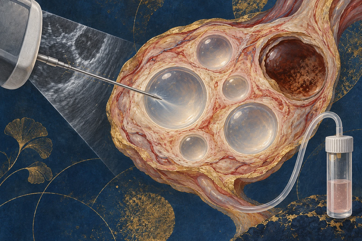
Endometriomas and retrieval difficulty
Endometriomas can affect access and specimen contents. Adhesions, a fixed ovary or a cyst obstructing genuine follicles may require a different route, sometimes through the cyst. Once cyst contents enter the needle and tube, aspiration can slow and specimen assessment may become harder. A prospective study of 56 women with endometriomas and 227 controls reported cyst transfixion in 14%, contamination in 16%, and an odds ratio of 3.6 for incomplete follicular aspiration. [6]
The same study found no significant increase in subjectively assessed overall procedural difficulty and no significant difference in oocytes recovered per developed follicle. The authors described increased difficulties of limited overall magnitude. When suspected endometrioma and low yield coexist, review lesion identity, needle access and individual tube contents. Post-procedure descriptions of fluid resembling endometrioma contents are particularly dependent on color, collection sequence and the preoperative ultrasound. [6]

Other stages that can reduce yield
Partial premature ovulation can lower oocyte yield. A retrospective comparison with matched controls found approximately 6.1 versus 11.2 retrieved oocytes, with fewer mature oocytes but without corresponding reductions in the maturity and fertilization rates of those recovered. This supports a quantity effect when some follicles have already ruptured; it does not establish that premature ovulation makes all aspirate viscous or repeatedly blocks needles. Relevant findings include hormones around triggering, follicular collapse, pelvic free fluid and actual retrieval timing. A weakly positive urine ovulation test alone does not establish inadequate antagonist suppression. During review of unusually low yield, the clinical team can assess medication, test timing, serum LH and progesterone trends. [8]
Premature luteinization is another possible change before retrieval. Follicles can acquire a luteal state without rupturing when LH action occurs early without a fully ovulatory response. A 1987 ultrasound study of 271 stimulated patients observed wall thickening and irregular internal echoes in 21. Eight had a preceding clear LH peak and subsequent progesterone rise; the other 13 had a smaller LH increase or medication-related changes. Progesterone was higher before triggering and at retrieval. During corpus luteum formation, vessels enter the inner follicular wall and blood may enter the cavity. This offers a physiological explanation for bloody, viscous aspirate, although dedicated measurements of such samples are lacking. Luteinized follicles can retain a measurable diameter rather than collapsing, unlike ovulated follicles; Figure 3 compares these states. [21]
A 2026 case report described premature luteinization confined to large follicles in one ovary, with smaller follicles in the other unaffected. Repeated late-follicular serum measurements showed an abnormal progesterone rise without a premature LH rise. The authors proposed that mechanical pressure associated with large follicles might contribute. A single case supplies no incidence estimate. If bloody viscous fluid is concentrated in the largest follicles while medium-sized follicles yield oocytes normally, review trigger-day size distribution and pre-trigger LH, progesterone and estradiol. [22]
Follicle size and mature-oocyte yield are associated over ranges. An analysis of 499 cycles found trigger-day follicles of 12–19 mm contributed most to retrieved and mature oocytes. A multicenter study of 19,082 patients identified 13–18 mm and associated a higher proportion above 18 mm with an early late-stimulation progesterone rise. These are population findings. A study using retrieval-day diameters found good yield from follicles measuring 19–24.5 mm. Trigger timing therefore balances leading and smaller follicles, with possible costs to prolonged waiting. [23,24,25]

Trigger administration, absorption and timing also warrant review when yield is unusually low. ESHRE advises considering trigger effectiveness promptly if the earliest tubes contain unusually clear fluid with almost no granulosa or cumulus cells and no oocytes, checking appropriate markers for the trigger used. This is an investigation pathway for nearly zero yield. Recovery of mature oocytes does not fit the typical picture of complete trigger failure, although timing and administration records remain relevant. A low count alone does not establish genuine empty follicle syndrome. [1,20]
Ovarian reserve and stimulation response inform expected yield at population level. A retrospective study of 10,624 cycles defined unsatisfactory recovery as fewer oocytes than trigger-day follicles larger than 14 mm: 1,294 cycles, approximately 12.2%, met this definition. Age, baseline FSH and endometriosis were associated factors. The study did not specifically select bloody viscous fluid with needle blockage or quantify responsibility attributable to patients and clinicians. ASRM considers AMH and antral follicle count useful for predicting stimulation-related quantity, with weak predictive ability for oocyte quality and live birth. Age or one AMH measurement alone cannot explain documented needle obstruction. [9,12]
This cohort's definition also matters statistically. Comparing oocyte yield with trigger-day follicles above 14 mm measures a broad quantity discrepancy, potentially involving maturity, natural variation, pathology, procedural factors and laboratory assessment. Associations with endometriosis or older age do not identify the cause of a particular obstructed needle or bloody sample. [9]
The final stage is laboratory identification. ESHRE recommends examining follicular fluid under a stereomicroscope in warmed conditions, documenting retrieval time, count, operator and witness. A 2026 four-center study reprocessed fluid already searched manually and designated for disposal using a microfluidic prototype. At least one additional oocyte was recovered in 316 of 582 patients. The findings identify opportunities to improve recovery, but the device is not universal routine equipment and the study cannot establish that a particular operation necessarily missed oocytes. Bloody or debris-rich specimens still require careful searching under the center's method and real-time communication with the retrieval clinician. [2,16]
Clinical management of needle blockage
ESHRE addresses sudden suction failure as an intraoperative troubleshooting task. Maintain visualization of the needle tip and check the suction pump, connections, kinked tubing and tube seal. Under ultrasound, assess follicular collapse and whether the wall covers the opening. Slight rotation may release wall obstruction while the needle remains in the follicle. If aspiration does not resume, withdraw and backflush with culture medium, then aspirate a small amount of medium outside the body to confirm patency before re-entry. A double-lumen needle can permit flushing without leaving the ovary under specified conditions. Replace the needle if patency cannot be restored. After entering an endometrioma or hemorrhagic follicle, replace the collection tube as well. [1]
The aim is to restore verifiable flow and check the remaining follicles. One or more needle replacements alone establish neither error nor completion of every recommended step. Records should specify the site and time of failure, fluid color, whether flushing or replacement restored flow, and which subsequent follicles were aspirated with observed collapse. The laboratory reports oocytes and granulosa cells concurrently, and the team checks for intraovarian or extraovarian bleeding before completing the procedure. [1,2]
Backflushing a blocked system and routinely repeatedly flushing every follicle serve different purposes. Pooled randomized trials have not shown that routine follicular flushing improves live birth and indicate longer procedure times. Routine additional flushing of all follicles is therefore not a general remedy for a yield discrepancy. [15]
When several consecutive tubes contain no oocytes, the laboratory's description is particularly important: granulosa cells without oocytes, nearly cell-free clear fluid, or extensive clots and debris suggest different investigations. ESHRE's trigger-check pathway relies on signals from the earliest tubes and clinical assessment of any immediate testing or change in strategy. Incomplete aspiration and difficult specimen assessment also require consideration. [1]
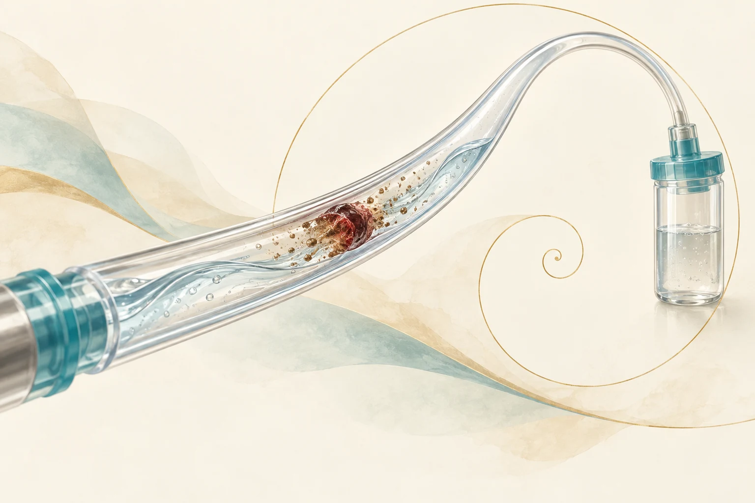
Figure 2 · Clinical checks when aspiration fails
- Check the systemSuction pump, connections, kinked tubing and tube seal.
- Check the tip and follicleContinuous ultrasound observation; assess wall obstruction after collapse.
- Release obstructionMinor adjustment according to guidance; withdraw and backflush with culture medium when needed.
- Verify before continuingReplace an unpatent needle and confirm flow outside the body before re-entry.
- Complete tube-specific checksReal-time laboratory counts; review remaining follicles and bleeding.
Based on ESHRE 2019 retrieval guidance. Specific actions are determined by the clinical team.
Assessing patient conditions, procedure and stimulation decisions
Documented patient conditions can create difficulty: an imaging-confirmed endometrioma obstructing access, adhesions fixing the ovary, premature rupture of some follicles, or a confirmed bleeding tendency. Each can change access, fluid composition or the time window and needs independent evidence. Bloody fluid, viscosity and needle obstruction alone cannot identify a disease or justify a general claim that patient factors usually predominate. [1,4,6,8,18]
Procedural quality is also assessable: cyst recognition and route planning, continuous tip visualization, confirmation of collapse, restoration of aspiration after blockage, completion of safely accessible follicles, and tube-by-tube laboratory feedback. Retrieval has a learning curve; a prospective training study found substantial differences between trainees in the procedures needed to reach predefined proficiency. Technique can affect results, while one low-yield procedure alone cannot establish a specific clinician's fault. [1,13]
Stimulation and trigger decisions are a third area. Follicular state is largely established by the time the retrieval clinician takes over. A 1997 dose study found daily cetrorelix 0.25 mg effective against a premature LH surge in its controlled-stimulation protocol. A 2008 clomiphene/HMG stimulation study for intrauterine insemination observed premature LH surges in 18.9–21.6% of patients despite cetrorelix 0.25–0.5 mg daily. That proportion belongs to its specific protocol and population; the treating team determines actual medication. Late-follicular hormone measurements, leading-follicle size at triggering and changes in monitoring provider can also be reviewed. These records help locate possible problems rather than independently establish fault. [26,27]
Factors can overlap. Leading follicles may change before triggering and produce bloody viscous fluid; a cyst may narrow access; puncture bleeding may add clots; and prolonged troubleshooting may leave later follicles incompletely aspirated. Follicular state and pathology can initiate difficulty, while recognition and troubleshooting influence its consequences. Assessment should reconstruct a verifiable timeline rather than assign predetermined shares of responsibility.

Confirmed endometriomas, substantial adhesions and abnormal ovarian position primarily affect access and contamination risk. Partial premature ovulation affects how many oocytes remain within follicles. Bleeding disorders and anticoagulants primarily affect bleeding risk. Age and diminished reserve affect average expected yield. These conditions can coexist while acting at different stages, so a complete account of repeated bloody tubes, blockage and unexpectedly low yield requires the process records.
Professional guidance has not established weight, stress, diet or ordinary menstrual discomfort as direct causes of this particular combination. Endometriosis also requires more evidence than dysmenorrhea or an ambiguous cyst image. The most discriminating synchronous records identify confirmed cysts, the onset of reduced flow, premature rupture and the fluid submitted from each ovary.
Table 1. Clinical clues and review directions. Clues do not replace pathology or complete intraoperative records.
| Clue | Stage potentially involved | Contemporaneous evidence needed |
|---|---|---|
| Typical cyst echoes before puncture; dark-brown thick first tube | Endometrioma or another cystic lesion in the access route | Complete ultrasound, Doppler, needle route and first-tube appearance |
| Initially clear fluid progressively turns red after puncture | Puncture bleeding or residual blood in tubing | Tissue traversed, tube sequence and changes after flushing |
| Some follicles have collapsed; pelvic free fluid | Partial premature ovulation | Trigger-to-retrieval interval, hormones and retrieval-day ultrasound |
| Medium follicles yield oocytes; the largest contain blood without oocytes | Changes in leading follicles or premature luteinization | Trigger-day size distribution and pre-trigger LH, progesterone and estradiol |
| Repeated loss of flow resolves after needle replacement | Wall obstruction, clots, tubing or equipment | Suction pressure, tip position, flushing response and collapse |
| Adequate specimen volume with few oocytes | Follicular maturity or laboratory-search review | Tube volumes, cells and laboratory records |
Improvements supported by the evidence
Persistent complex cystic structures should be assessed by a clinician experienced in reproductive ultrasound, including their spatial relationship to accessible follicles, internal echoes, vascularity and follow-up changes. For a confirmed endometrioma, a subsequent-cycle plan can specify avoidance, replacement of contaminated collection tubes and infection-risk discussion. ESHRE does not recommend routine endometrioma surgery solely to improve ART live birth because surgery may harm ovarian reserve. Substantial pain or impeded follicular access can support an individualized surgical discussion. [1,4,14]
When records support premature ovulation or luteinization, review LH-surge suppression, monitoring times, trigger decisions and the actual trigger-to-retrieval interval. Changes depend on subsequent growth and hormones rather than one universal diameter or retrieval one day earlier. ESHRE describes triggering commonly when several leading follicles reach 16–22 mm, with individualized timing; the cited analysis associated trigger-day 12–19 mm follicles with the greatest mature-oocyte contribution. Unequal growth requires balancing smaller and leading follicles. ESHRE does not require routine additional hormone testing in standard protocols. When leading follicles are unusually large, medication differs from standard practice or early LH changes are suspected, the team may evaluate serum LH, progesterone and ultrasound. Urine-strip results also require interpretation in light of test timing and medication. [8,19,23,24]
Figure 3 · Three states before retrieval
Clear cavity and maintained contour.
Clear or pale-yellow fluid; an oocyte may be identified.
Diameter remains measurable; wall thickening and internal echoes may appear.
Fluid may be bloody or viscous; recovery can be difficult.
Follicle collapses or disappears; pelvic free fluid may occur.
Little or no fluid; the oocyte has been released.
Different states may coexist in one cycle. Diameter alone does not distinguish maturation from luteinization.
If records indicate insufficient effective aspiration after blockage, include equipment checks, tip visibility, confirmation of restored flow and side- and tube-specific records in the next plan. Adequate submitted fluid with few identified oocytes calls for specimen-handling and laboratory-record review. The microfluidic study indicates a possible future aid, rather than a universally implemented standard rescue. [1,2,16]
A complete review needs at least six record groups: trigger medication and administration time; trigger- and retrieval-day ultrasound, hormones and pelvic free fluid; actually punctured follicles and aspirate volumes; blockage times and flushing or replacement; each tube's appearance and oocyte search; and postoperative bleeding assessment. Each maps a possible loss pathway and informs whether pathology, timing, access or laboratory workflow deserves priority. [1,2]
The retrieval clinician, embryology laboratory and stimulation clinician should review these together. Detailed records turn risks into practical arrangements: which structures to avoid, which follicles may be inaccessible, who alerts whom after the first abnormal tube, when to change tubes, when to request immediate laboratory feedback, and how to monitor postoperative bleeding. Sufficient detail allows the team to assess whether a subsequent change addressed the documented problem.

Needle clots and postoperative thrombosis
Clots within the retrieval system usually arise from coagulation of blood in the local aspirated specimen. They do not establish deep-vein thrombosis or a systemic hypercoagulable state. Thrombosis assessment after stimulation depends principally on previous thrombosis, ovarian hyperstimulation syndrome risk, symptoms and clinical examination. Unsupervised anticoagulant use can increase bleeding. Breathlessness, chest pain, unilateral leg swelling or pain, or rapidly worsening abdominal distension or pain warrant prompt medical assessment and possible emergency or specialist care. [17,18]
Conclusion
Bloody viscous aspirate and a blocked retrieval needle can arise through several mechanisms, as can low yield. Current evidence supports identifying cystic or hemorrhagic structures, troubleshooting blockage, verifying collapse and checking laboratory recovery. It does not quantify a general predominant patient responsibility for this combination. The most useful explanation aligns fluid source, failure timing, effective punctures and tube-specific results with the follicular state before triggering. Locating the loss guides the next-cycle review.
Scope and limitations
This article addresses a clinical phenomenon without presenting, inferring or evaluating any identifiable patient's outcome. Many procedural recommendations rely on expert consensus, and some mechanism studies are small. Associations and feasible emergency steps do not replace review of a particular cycle's original records. The illustrations are AI concepts and the explanatory diagrams are editorial drawings; none is a medical image.
Review retrieval using the original records
FS can help organize stimulation, trigger, retrieval and laboratory records. The treating team assesses the cause and any next-cycle changes using the complete information.
References and original guidance
Reference numbers match the text. Initial sources were checked on 27 September 2026, with references 21–27 added on 30 September. Key guidance and studies were reviewed for this page on 2 October.
- ESHRE Working Group on Ultrasound in ART. Recommendations for good practice in ultrasound: oocyte pick up. Hum Reprod Open. 2019;hoz025.
- ESHRE IVF labs working group. ESHRE recommendations on Good Practice in the IVF laboratory. Hum Reprod. 2026;deag096.
- ESHRE SIG Embryology and Alpha Scientists. Vienna consensus on ART laboratory performance indicators. Reprod Biomed Online. 2017.
- ACR O-RADS US Committee. O-RADS US v2022 update. Radiology. 2023.
- Catheter-directed sclerotherapy for ovarian endometrioma: short-term outcomes. Radiology. 2018.
- Benaglia L, et al. Oocyte retrieval difficulties in women with ovarian endometriomas. Reprod Biomed Online. 2018.
- Levine D, et al. Management of asymptomatic ovarian and other adnexal cysts imaged at US. Radiology. 2010.
- Detecting partial premature ovulation during follicular aspiration compromises quantity but not quality. 2024.
- Wang et al. Causes and Effects of Oocyte Retrieval Difficulties: 10,624 Cycles. Front Endocrinol. 2022.
- Fisch B, et al. Viscosity and refractive index of follicular fluid in relation to IVF. J Assist Reprod Genet. 1996.
- Follicular fluid rheology and the duration of the ovulatory process. Hum Reprod. 2000.
- ASRM Practice Committee. Testing and interpreting measures of ovarian reserve. Fertil Steril. 2020.
- Proficiency in oocyte retrieval assessed by the learning curve cumulative summation test. Fertil Steril. 2014.
- ESHRE Endometriosis Guideline Group. ESHRE guideline: endometriosis. Hum Reprod Open. 2022.
- Follicle flushing does not improve live birth and increases procedure time: systematic review and meta-analysis. Fertil Steril. 2021.
- Microfluidic automation improves oocyte recovery from follicular fluid. Nat Med. 2026.
- RCOG. Ovarian hyperstimulation syndrome patient information.
- Hoffman R, Brenner B. Corpus luteum hemorrhage in women with bleeding disorders. Womens Health. 2009.
- ESHRE guideline on ovarian stimulation for IVF ICSI. Update 2025.
- Empty follicle syndrome: a systematic review. Fertil Steril. 2008.
- Hamori M, et al. Premature luteinization of follicles during ovarian stimulation for in-vitro fertilization. Hum Reprod. 1987.
- Kazatsker MM, Kol S, Khoury S. Mechanical luteinization of follicles before oocyte retrieval is often overlooked: a case report. J Assist Reprod Genet. 2026.
- Abbara A, et al. Follicle size on day of trigger most likely to yield a mature oocyte. Front Endocrinol. 2018.
- Hanassab S, et al. Explainable artificial intelligence to identify follicles that optimize clinical outcomes during assisted conception. Nat Commun. 2025.
- Shapiro BS, et al. The effect of ovarian follicle size on oocyte and embryology outcomes. Fertil Steril. 2022.
- Albano C, et al. Comparison of different doses of gonadotropin-releasing hormone antagonist Cetrorelix during controlled ovarian hyperstimulation. Fertil Steril. 1997.
- Lin YH, et al. Effect of cetrorelix dose on premature LH surge during ovarian stimulation. Reprod Biomed Online. 2008.
For public medical education only; not individual medical advice.